T1 Hypointense Suprasellar Lesion
Anne G. Osborn, MD, FACR
DIFFERENTIAL DIAGNOSIS
Common
Dilated Third Ventricle
Arachnoid Cyst (AC)
Neurocysticercosis (NCC)
Less Common
Pilocytic Astrocytoma (PA)
Craniopharyngioma
Epidermoid Cyst
Rathke Cleft Cyst
Enlarged Perivascular Spaces
Rare but Important
Pituitary Macroadenoma
Saccular Aneurysm (Acutely Thrombosed)
Pituitary Apoplexy
Pilomyxoid Astrocytoma
ESSENTIAL INFORMATION
Key Differential Diagnosis Issues
T1 hypointense lesion = hypointense compared to brain, not necessarily ˜ CSF
Lesions CSF-like on all sequences
Enlarged 3rd ventricle, arachnoid cyst, perivascular spaces
Epidermoid cyst
Lesions hypointense to brain but hyperintense to CSF on T1WI
Neoplasms
Congenital or infectious cysts
Enhancing hypointense lesions
Solid (astrocytoma, adenoma)
Rim/ring (NCC, craniopharyngioma, RCC)
Helpful Clues for Common Diagnoses
Dilated Third Ventricle
Enlarged 3rd ventricle recesses protrude into suprasellar cistern, sella turcica
Behaves exactly like CSF on FLAIR, DWI
Arachnoid Cyst (AC)
Bows 3rd ventricle up, over cyst
Suppresses on FLAIR, no restriction on DWI
Neurocysticercosis (NCC)
Cysts often show rim enhancement
Helpful Clues for Less Common Diagnoses
Pilocytic Astrocytoma (PA)
Hyperintense to CSF on T1WI
Enhancement typical
Craniopharyngioma
90% cystic, 90% Ca++, 90% enhance
Cyst signal variable (hyper > hypointense)
Epidermoid Cyst
No suppression on FLAIR, restricts on DWI
Rathke Cleft Cyst
Cyst fluid more often hyperintense
Look for “claw sign” of enhancing pituitary around cyst
Helpful Clues for Rare Diagnoses
Pituitary Macroadenoma
Small intratumoral cysts common
Extratumoral cysts (trapped perivascular spaces) less common
Necrosis/apoplexy may appear cystic, show rim enhancement
Image Gallery
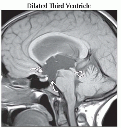 Sagittal T1WI MR shows aqueductal stenosis
 causing marked enlargement of the third ventricle with herniation of anterior recesses into the suprasellar cistern causing marked enlargement of the third ventricle with herniation of anterior recesses into the suprasellar cistern  . .Related posts:Stay updated, free articles. Join our Telegram channel
Full access? Get Clinical Tree
 Get Clinical Tree app for offline access

|