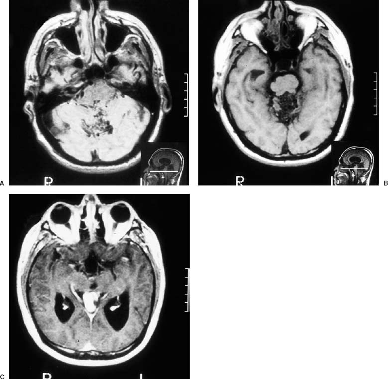
27 Diagnosis High-flow cerebral arteriovenous malformation of the cerebellar vermis Problems and Tactics A patient with a relatively large, high-flow arteriovenous malformation (AVM) was operated for complete excision of his AVM in one stage without preoperative embolization. The operation went well, but the patient did not wake up and remained in a coma for several days with very slow and gradual improvement over the next several months. The postoperative studies showed clearly that he had suffered extensive retrograde thrombosis of the entire deep venous system that had previously drained the AVM. It is possible that this complication could have been prevented by “staged” treatment of the AVM. Keywords Cerebral arteriovenous malformation, venous occlusion, retrograde venous thrombosis, embolization This 35-year-old, healthy man presented with trigeminal neuralgia of several years’ duration. His workup showed a large, high-flow AVM of the cerebellar vermis (Figs. 27–1 and 27–2). It was thought that the trigeminal neuralgia was probably secondary to venous compression of the trigeminal nerve by arterialized veins. His AVM was excised surgically in a one-stage operation. He remained in a coma after surgery and a computed tomographic (CT) scan showed that the surgical bed was dry, but there was a suggestion of thrombosis within the basal vein of Rosenthal bilaterally (Fig. 27–3). A postoperative arteriogram showed complete removal of the malformation and extensive venous thrombosis with essentially no filling of the entire deep venous system (Fig. 27–4). He was treated initially with hydration and then later with anticoagulation, but he remained in a coma, although his cranial nerves functioned normally. He had moderate hydro-cephalus preoperatively, and although the ventricles did not increase in size postoperatively, we treated him with a ventriculostomy and later with a VP shunt, but he showed no improvement initially. Very gradually, he began to improve to the point that several months later, he was able to walk slowly and move all his limbs well, although he still had significant spasticity of his arms and legs. His cognitive function had recovered to his preoperative status. A recent CT scan does not show any significant infarction (Fig. 27–5). FIGURE 27–1 (A,B) Preoperative magnetic resonance imaging showing large, but compact arteriovenous malformation of the anterior superior cerebellar vermis. (C) Note the very distended vein of Galen.
Retrograde Venous Thrombosis after Excision of a Cerebral Arteriovenous Malformation
Clinical Presentation
Surgical Technique
Related posts:
 28 Dominant Hemisphere Insular Tumor
 9 Large Aneurysm of the Internal Carotid Artery Obliterated with Seven Fenestrated Clips under Intraoperative Monitoring of Anterior Choroidal Arterial Blood Flow Insufficiency
 20 Collateralization via Vasa Vasorum: A Determinant of Therapeutic Efficacy of Coil Embolization of the Thrombosed Giant Aneurysm of the Vertebral Artery
 19 Partially Thrombosed Giant Dissecting Aneurysm of the Vertebral Artery—Treatment Strategies
 18 Combined Endovascular and Surgical Treatment of a Large Basilar Trunk Aneurysm in a Child
 62 Intramedullary Spinal Arteriovenous Malformation
Stay updated, free articles. Join our Telegram channel

Full access? Get Clinical Tree


