Vignette 3
The Rapid Eye Movement Sleep Behavior Disorder Leading to a Subdural Hemorrhage
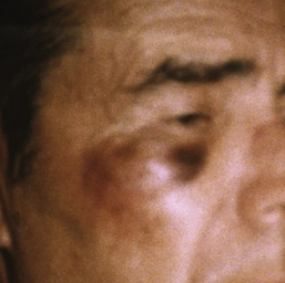
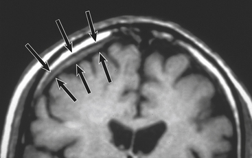
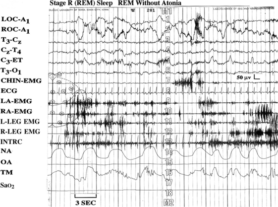
A 73-year-old man had 10 years of progressively more frequent, almost nightly dreams in which “I act them out, sometimes I hurt myself, I go through the motions of what I’m dreaming about.”1,2 He recently dreamed about a man running; “Someone yelled, ‘Stop him.’ I tried.” His wife reported that immediately before awakening he jumped from the bed and sustained superficial injuries. During another event he struck the right side of his face, then complained of a headache, vomited, and appeared confused. His examination at that time showed a right infraorbital hematoma (Vignette Fig. 3.1). A magnetic resonance imaging scan of the brain and brainstem showed a right subdural hematoma (Vignette Fig. 3.2). Standard polysomnography (PSG), using split-screen electroencephalographic video analysis, revealed significant periodic limb movements (with a movement index of 52 events per hour) and an unusual increase in phasic limb movements throughout most of the rapid eye movement (REM) sleep periods, or “REM sleep without atonia” (Vignette Fig. 3.3). During one REM sleep episode, after a relatively prolonged period of uncharacteristically quiet electromyographic activity, the patient (a cattle farmer) suddenly exhibited explosive running movements, after which he awoke and, in an alert and coherent manner, spontaneously reported “one of them dreams—I was chasing cattle” (Vignette Fig. 3.4).

VIGNETTE FIGURE 3.1 The patient at the time of his initial examination had evidence of superficial injury sustained to the right maxillary region from a reported episode of violent sleep-related behavior. (From Dyken ME, Lin-Dyken DC, Seaba P, et al. Violent sleep-related behavior leading to subdural hemorrhage. Arch Neurol. 1995;52:318-321.)

VIGNETTE FIGURE 3.2 A brain magnetic resonance image of the patient in Vignette Figure 3.1 revealed a subdural hemorrhage, as outlined by the black arrows. (From Dyken ME, Lin-Dyken DC, Seaba P, et al. Violent sleep-related behavior leading to subdural hemorrhage. Arch Neurol. 1995;52:318-321.)

VIGNETTE FIGURE 3.3 The patient’s polysomnographic tracing revealed an unusually high level of electromyographic (EMG) augmentation during rapid eye movement sleep. LOC, Left outer canthus; ROC, right outer canthus; T, temporal; C, central; ET, ears tied; O, occipital; LA, left arm; RA, right arm; L-LEG, left leg; R-LEG, right leg; INTRC, intercostal EMG; NA, nasal airflow; OA, oral airflow; TM, thoracic movement. (From Dyken ME, Yamada T, Lin-Dyken DC. Polysomnographic assessment of spells in sleep: nocturnal seizures versus parasomnias. Semin Neurol. 2001;21[4]:377-390.)


