A New Cervical Disc Prosthesis: Mobi-C. Preliminary Results of a Prospective Study
Pierre Bernard
Jean-Marc Vital
Thierry Dufour
Jacques Beaurain
Jean-Marc Fuentes
Jean Huppert
Istvan Hovorka
With most joints, for several decades now, arthrodesis has already been superseded by prosthesis implantation. In view of the good results of the decompression-arthrodesis process, the vertebral column was left out of this trend for a very long time, until modern and efficient prostheses became available in the 1980s for lumbar discs and 10 to 15 years later for cervical discs. At the same time, more and more studies have shown the harmful effect of cervical intervertebral fusion on the adjacent levels, which tend to degenerate more rapidly. This argument encouraged the development of several interbody implants allowing preservation of intervertebral mobility, thus avoiding overstressing neighboring segments. The Mobi-C prosthesis has been developed with this perspective, giving particular care to simplifying ancillary equipment and surgical technique.
Rationale for Cervical Disc Arthroplasty
At the beginning of the 1950s, Cloward (6) and Smith and Robinson (24) described the anterolateral approach route to the cervical spine and its use in the treatment of compressive disc disease. The performance of an isolated discectomy without associated fusion is recognized as being generally deceptive, as this technique can lead to new cervicalgia caused by postdiscectomy instability and also secondary local kyphosis. Over the years, this anterior surgery has been complemented with plate osteosynthesis then interbody fusion cage, and, increasingly, the use of nonautologous intervertebral grafts: bone substitutes or even allografts replacing bone graft of iliac origin. On the whole, these operations give a highly satisfactory and durable result with regard to both cervical and brachial pain, while retaining generally satisfactory cervical function and mobility, often compensated by neighboring levels. Therefore, the decompression-arthrodesis combination is currently the technique of reference for most cervical pathologies requiring surgical intervention. However, in a large number of cases the initial symptom that gives rise to surgery is neurologic, and complementary arthrodesis is only carried out to prevent the possibility of iatrogenic instability of the total discectomy. This fusion can lead to restricted cervical mobility. Moreover, this operation is not without disadvantages: risk of pseudarthrosis, morbidity associated with harvesting an iliac graft if this is required, and so forth.
At the same time more and more evidence has accumulated concerning the repercussions of arthrodesis on the adjacent cervical levels by acceleration of the degeneration
phenomena. These arguments are at the same time biomechanical, clinical, and radiologic. Eck et al. (10), DiAngelo et al. (9), Wigfield et al. (26), and Matsunaga et al. (20) showed in biomechanical studies an increase in stresses and mobility of a disc segment situated beside a fused area. Many clinical studies have also shown the same. Hilibrand et al. (16) reviewed 409 cervical arthrodeses in 374 patients with a follow-up of up to 21 years. They reported an incidence of occurrence of symptomatic degeneration of an adjacent level of 2.9% per year in the first 10 years, projecting that about a quarter of the patients operated on would suffer another disc problem in the first decade. In their group of patients, more than two thirds underwent a further operation. Gore and Sepic (14) reported that 16% of the 50 patients operated on and monitored with an average follow-up of more than 20 years had to undergo further operations on another level. In their group of patients, Goffin et al. (12) found discopathy on an adjacent level in 92% of cases with a follow-up of only 5 years. Finally, several radiologic studies (2,17,25) showed accelerated wear occurring in the subsequent years above or below a cervical arthrodesis. All these arguments have weighed against the development of cervical arthroplasty. In addition to sparing the adjacent levels, an artificial cervical disc must enable the physiologic local lordosis to be regained, intervertebral mobility to be retained, and the postoperative immobilization, which is usually the rule following arthrodesis, to be dispensed with. Finally, it eliminates the disadvantages associated with harvesting an iliac graft.
phenomena. These arguments are at the same time biomechanical, clinical, and radiologic. Eck et al. (10), DiAngelo et al. (9), Wigfield et al. (26), and Matsunaga et al. (20) showed in biomechanical studies an increase in stresses and mobility of a disc segment situated beside a fused area. Many clinical studies have also shown the same. Hilibrand et al. (16) reviewed 409 cervical arthrodeses in 374 patients with a follow-up of up to 21 years. They reported an incidence of occurrence of symptomatic degeneration of an adjacent level of 2.9% per year in the first 10 years, projecting that about a quarter of the patients operated on would suffer another disc problem in the first decade. In their group of patients, more than two thirds underwent a further operation. Gore and Sepic (14) reported that 16% of the 50 patients operated on and monitored with an average follow-up of more than 20 years had to undergo further operations on another level. In their group of patients, Goffin et al. (12) found discopathy on an adjacent level in 92% of cases with a follow-up of only 5 years. Finally, several radiologic studies (2,17,25) showed accelerated wear occurring in the subsequent years above or below a cervical arthrodesis. All these arguments have weighed against the development of cervical arthroplasty. In addition to sparing the adjacent levels, an artificial cervical disc must enable the physiologic local lordosis to be regained, intervertebral mobility to be retained, and the postoperative immobilization, which is usually the rule following arthrodesis, to be dispensed with. Finally, it eliminates the disadvantages associated with harvesting an iliac graft.
The Mobi-C Prosthesis
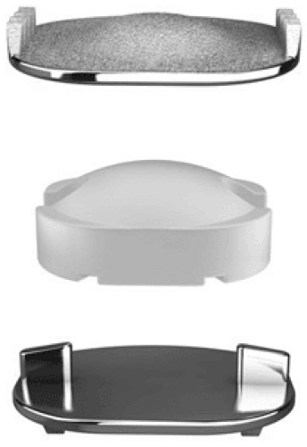
The Mobi-C artificial disc (LDR médical, Troyes, France) is made up of two vertebral plates (one superior and one inferior) and a polyethylene mobile insert (Fig. 29.1). Different sizes are available (13 ÷ 15, 13 ÷ 17, 15 ÷ 17 and 15 ÷ 20, depth ÷ length). The plates are manufactured from cobalt-chrome, and the surface in contact with the vertebral body has a coating of plasma sprayed porous titanium to facilitate bone integration. Different insert heights are available to restore the physiologic height of the disc (5, 6, and 7 mm). The self-positioning of the superior plate versus the inferior plate, through the controlled mobility of the inlay permits a uniform and more physiologic load distribution on the discovertebral segment. The “non-constraining” nature of this disc prosthesis limits the constraints on the bone-implant interface and favors the decrease of the constraints on the posterior facet joints. The self-centering of the mobile insert favors the respect of the instantaneous rotation centers and also gives back to the treated intervertebral segment its natural physiologic movements within the respect of the cervical lordosis.
To implant this prosthesis, the surgical approach is identical to a classical anterior cervical arthrodesis. Patients will first undergo conventional anterior discectomy to remove the dam-aged disc. When the disc is largely exposed, the midline of the vertebra is located thanks to the width gauge, and a radio-opaque centering pin is placed on this midline at the inferior edge of the superior plate or at the upper edge of the inferior plate of the vertebrae of the operated level. It is used as a reference mark throughout the surgery to be certain of the accurate
centering of the prosthesis. The intersomatic space is then distracted by distraction forceps and distraction is then maintained by a Caspar distractor. Total discectomy is then completed, up to the posterior ligament. The vertebral endplates are cleaned to remove the osteophytes and to make the flattest surface possible on the inferior plate. The aim is to be able to push the prosthesis to the posterior limit of the disc space. With the disc perfectly cleaned, one may proceed with the choice of the device. The depth gauge allows determination of the appropriate template size use. The distractor allows obtaining an intervertebral space of the desired height, and one proceed then to introduction of the trial implant corresponding to the size and the height of the future prosthesis. This trial implant must be inserted to the maximum depth because its position will dictate the correct placement of the prosthesis.
centering of the prosthesis. The intersomatic space is then distracted by distraction forceps and distraction is then maintained by a Caspar distractor. Total discectomy is then completed, up to the posterior ligament. The vertebral endplates are cleaned to remove the osteophytes and to make the flattest surface possible on the inferior plate. The aim is to be able to push the prosthesis to the posterior limit of the disc space. With the disc perfectly cleaned, one may proceed with the choice of the device. The depth gauge allows determination of the appropriate template size use. The distractor allows obtaining an intervertebral space of the desired height, and one proceed then to introduction of the trial implant corresponding to the size and the height of the future prosthesis. This trial implant must be inserted to the maximum depth because its position will dictate the correct placement of the prosthesis.
Stay updated, free articles. Join our Telegram channel

Full access? Get Clinical Tree


