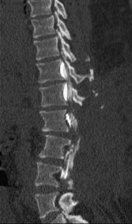
84 A 46-year-old achondroplastic dwarf came to the emergency department with cauda equina syndrome. A sagittal view, reconstructed computed tomography (CT) myelogram of the lumbar spine is seen, notable for the dysmorphic vertebral bodies and severe congenital spinal canal stenosis seen in this disorder (Fig. 84-1). Spinal stenosis in an achondroplastic dwarf An emergent lumbar laminectomy was done. There are hundreds of types of dwarfism, and achondroplasia is the most common. Achondroplastic dwarfs have short arms and legs, an enlarged head, and an average size trunk. They are about 4 feet in height. Abnormal endochondral ossification is the primary defect. This disorder has an autosomal-dominant inheritance, but ~80% of cases are due to a new mutation. Intelligence is normal. The most common health problems faced by this population are craniovertebral junction anomalies, lumbar stenosis, otitis media, sleep apnea, and obesity. The spine in achondroplastic children prior to ambulation has a thoracolumbar kyphosis, but corrects to an exaggerated lumbar lordosis with the development of ambulation. This spinal deformity is associated with hip flexion contractures and genu varum. Spinal stenosis can be severe in these patients. The pedicles and laminae are short and thick, and there is interpedicular narrowing, associated disc herniations, and vertebral body wedging.
Achondroplasia
Presentation
Radiologic Findings
Diagnosis
Treatment
Discussion
Achondroplasia
Only gold members can continue reading. Log In or Register to continue


Full access? Get Clinical Tree


