Fig. 1
Brain water content at 24 and 72 h post SBI. Brain water content was significantly higher in the ipsilateral frontal lobe (RF) of SBI animals than in shams at 24 h (a) and 72 h (b) post injury. Low-dose valproic acid (VA) pretreatment significantly reduced the brain water content increase within RF compared with vehicle-pretreated SBI animals at 24 h (a) post injury but not at 72 h (b). **p < 0.01 vs sham, ## p < 0.01 vs low-dose VA pretreatment, &p < 0.05 vs low-dose VA pretreatment. RF right frontal lobe, LF left frontal lobe, RP right parietal lobe, LP left parietal lobe, CB cerebellum, BS brain stem
At 24 h post surgery, VA pretreatment at the low dose significantly reduced brain edema in the right frontal lobe (81.88 ± 1.12 %) when compared with the vehicle-pretreated SBI (83.73 ± 1.21 %) and high-dose VA pretreatment SBI (83.30 ± 0.3 %). The high-dose pretreatment group did not significantly decrease right frontal lobe edema compared with vehicle-pretreated SBI (Fig. 1a).
At 72 h post surgery, there was a trend that low-dose VA pretreatment reduced brain water content within the right frontal lobe in comparison with the vehicle-pretreated SBI group (82.57 ± 1.1 % vs 83.32 ± 1.05 %), but this was not statistically significant (Fig. 1b).
MMP Enzymatic Activity in Brain after SBI
At 24 h post surgery, MMP-9 significantly increased in vehicle-pretreated SBI animals compared with shams (Fig. 2). However, pretreatment with low-dose VA did not significantly reduce the elevation of MMP-9 enzymatic activity. There was no significant difference in MMP-2 activity among sham, vehicle-pretreated SBI, and low-dose VA-pretreated SBI groups.
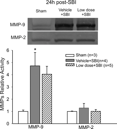
Fig. 2
MMP activities at 24 h post SBI. There was significantly increased MMP-9 but not MMP-2 activity in vehicle-pretreated SBI animals compared with sham. Low-dose valproic acid pretreatment did not significantly attenuate the elevation. *p < 0.05 vs sham
Neurobehavioral Deficits after SBI
At 24 and 72 h post surgery, modified Garcia and beam balance scores were significantly less in all SBI animals than shams, suggesting SBI-induced neurological deficit. Neither the low dose nor high dose of VA pretreatment significantly improved the impaired neurological function compared with vehicle-pretreated SBI (Figs. 3 and 4).
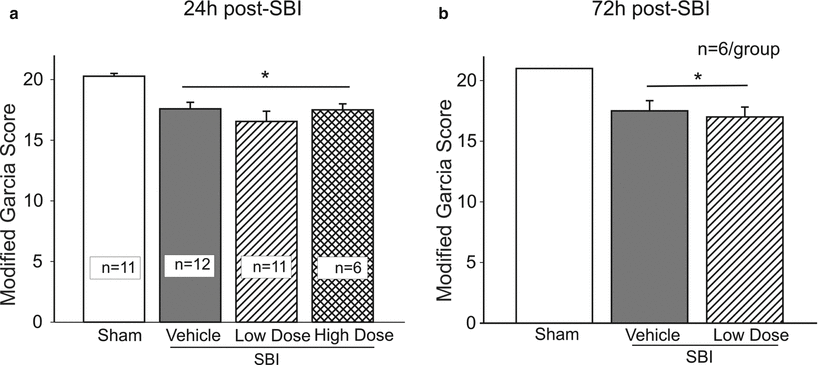
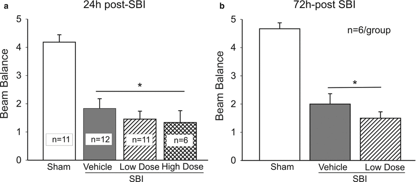
Fig. 3
Modified Garcia test at 24 and 72 h post SBI. SBI animals were associated with lower Garcia scores than shams at 24 h (a) and 72 h (b) post injury. Valproic acid pretreatment did not significantly improve Garcia scores at either time point. *p < 0.05 vs sham
Fig. 4
Beam balance test at 24 and 72 h post SBI. SBI animals were associated with lower beam balance scores than shams at 24 h (a) and 72 h (b) post injury. Valproic acid pretreatment did not significantly improve beam balance performance at either time point. *p < 0.05 vs sham
Discussion
In the present study, we demonstrated the neuroprotective effect of single-dose (100 mg/kg) VA against brain edema in a rat model of SBI. Our findings were that (1) VA pretreatment significantly attenuated brain edema at 24 h after SBI; (2) there were not significant suppressions of MMP activities associated with VA pretreatment; and (3) neurological deficits following SBI were not significantly improved by a single dose of VA pretreatment.
A rodent model of SBI introduced by our group has been shown to mimic the clinical human neurosurgery situation in that it results in the most typical postoperative consequences [7, 20]. It offers us a clinically relevant platform to study the basic pathophysiological mechanisms of SBI and to evaluate specific treatment strategies. In the right frontal lobe where brain tissue surrounds the resection site, there was a significant 3.3 % increase in water content compared with sham. Brain water content is a good indicator of brain swelling resulting from the edema. A 1 % increase in brain water content, equivalent to approximately a 4.3 % increase in tissue swelling, can lead to intracranial pressure elevation [9]. Our finding suggested the important role of brain edema associated with BBB disruption in the pathophysiology of SBI [7, 10, 20]. Neuroprotective strategies targeting brain edema would promise to limit postoperative complications; enabling more aggressive surgical approaches. The advantages and feasibility of preoperative treatment strategies rather than postoperative interventions for planned neurosurgical procedures have not been fully realized. In the present study, we demonstrated the efficacy of VA pretreatment against brain edema induced by SBI. When VA was administered 30 min before brain resection, it significantly reduced brain edema compared with vehicle-pretreated animals at 24 h after SBI. This result echoes studies in both transient and permanent middle cerebral artery occlusion (MCAO) models that show VA pretreatment attenuated ischemic brain damage and brain edema associated with BBB disruption [12, 18]. Similarly, 30 min post-injury administration of VA has also been shown to improve BBB integrity in a rodent model of traumatic brain injury [4]. The lack of protection persistent to 72 h implies that enhanced long-term neuroprotective effects may be achieved by multiple VA interventions before and after SBI.
MMPs are proteases that cleave the extracellular matrix, causing disruption of BBB, and play a critical role in brain injury associated with a variety of brain diseases [2, 3, 5]. In the current study, there was significantly higher MMP-9 activity at 24 h after SBI. It was consistent with our findings earlier using the SBI model that MMP-9 enzymatic activity was greatly elevated in the vulnerable peri-resection brain regions over the first 3 days post surgery [20]. VA pretreatment was associated with a lower level of MMP-9 activity, but this was not significantly different from that of vehicle-pretreated animals. The statistical insignificance may the result of the small sample size. On the other hand, it is possible that anti-inflammation and antioxidative effects also constitute the neuroprotective mechanisms of VA [17]. Following traumatic brain injury, BBB protection by VA may involve HDAC inhibition-mediated suppression of NF-κB and tight junction degradation in addition to MMP-9 suppression [18]. Additional research will help elucidate the underlying mechanism of VA against brain edema in the setting of SBI.
Related posts:
 of Behavioral Deficits in Rodents Following Brain Injury Across Species, Gender, and Experimental Model
 Infarction After Aneurysmal Subarachnoid Hemorrhage
 Volume Determination in Subarachnoid Hemorrhage Using Rats
 Endothelial Growth Factor in Brain Edema Formation After Subarachnoid Hemorrhage
 in Brain Swelling and Infarction Volume over Four Days After Hypoxia Ischemia in Neonatal Rats
 of Gender on Iron-induced Brain Injury in Low Aerobic Capacity Rats
Stay updated, free articles. Join our Telegram channel

Full access? Get Clinical Tree


