♦ Preoperative
Operative Planning
- Radiosurgery should be considered for small or medium sized tumors, depending on the patient’s age, medical conditions, hearing status, and personal preference
- Magnetic resonance imaging: determine location, size, and relationships of tumor to adjacent structures such as brain stem and vascular structures, including location of the jugular bulb, transverse, and sigmoid sinuses
- Computed tomography: assess bony anatomy of petrous temporal bone, middle fossa, posterior fossa, and encased arteries
Additional Testing
- Comprehensive audiologic evaluation: pure-tone audiometry, speech discrimination testing, brainstem auditory evoked responses in patients who cannot cooperate with routine assessments
Equipment
- Irrigating bipolar cautery
- Kartush dissector/nerve stimulator
- Ultrasonic aspirator
Anesthetic Issues
- Anesthesiologist needs to be aware that electrophysiological monitoring of cranial nerves will be performed.
♦ Intraoperative
Lateral Suboccipital (Retrosigmoid) Approach (see Chapter 14, Retrosigmoid Approach)
Pros
- Hearing preservation surgery
- Minimizes petrous bone drilling
Cons
- Requires cerebellar retraction
- Facial nerve typically away from surgeon upon initial approach
Removal of Small Tumors (Fig. 49.1)
- The inferolateral cerebellar hemisphere is retracted gently to allow cerebrospinal fluid (CSF) drainage from the cisterna magna.
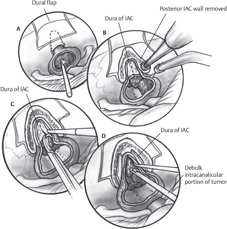
Fig. 49.1 (A–D) After removal of the posterior internal auditory canal, the intracanalicular portion of the tumor is debulked. IAC, internal auditory canal.
- Surgical dissection and resection are assisted by electrophysiologic monitoring of cranial nerves (CN) V, VII, and VIII for small to medium tumors as well as CN IX, X, and XI for larger tumors.
- After placement of the brain retractor on the lateral cerebellar hemisphere, the operating microscope is positioned.
- The facial and vestibulocochlear nerves, which are usually anteriorly displaced by the tumor, are identified both at the brain stem and within the internal auditory canal (IAC).
- Stimulation of the tumor capsule to determine aberrant course of facial nerve is mandated prior to cauterization and decompression .
- The facial nerve origin at the brain stem is adjacent to the pontomedullary sulcus, which marks the junction of the pons and medulla.
- The vestibulocochlear nerve joins the brain stem 1 to 2 mm posterior to the facial nerve origin.
- The origin of CN IX, X, and XI are 2 to 3 mm inferior to the origin of the facial nerve.
- After originating at the basilar artery, the anterior inferior cerebellar artery (AICA) courses laterally to supply branches to the cochlear nerve in the IAC, then forms a meatal loop that continues medially between CN VIII and CN IX to supply the brain stem and cerebellum.
- The meatal loop is usually situated in front of the porous acusticus or within the IAC.
- The AICA can be displaced inferiorly, anteriorly, or superiorly by the tumor.
- The posterior inferior cerebellar artery (PICA) originates from the vertebral artery in close proximity to CN IX, X, and XI.
- The petrosal vein is identified.
- The dura overlying the posterior wall of the acoustic meatus is coagulated, incised sharply with a no. 11 blade, and then mobilized with a curette.
- A high speed drill with a 5-mm and 2-mm diamond burr is used to remove the posterior wall of the IAC; the extent of the bone removal is guided by the size of the intracanalicular portion of the tumor.
- After the bone removal is completed, the intracanalicular dura is exposed, coagulated, opened horizontally, and reflected to reveal the intracanalicular part of the tumor, which has usually displaced the facial and cochlear nerves anteriorly.
- The arachnoid overlying the tumor is opened.
- The tumor capsule is coagulated and incised with microscissors.
- Internal decompression of the tumor is performed with suction, tumor forceps, and microfreer.
- Tumor dissection in the arachnoid plane outside the tumor capsule is performed with judicious use of the Kartush nerve stimulator dissector to confirm position of the facial nerve.
- Beginning at the medial boundary of the tumor, dissection along the facial and cochlear nerves proceeds from a medial to lateral direction by means of straight or curved microdissectors and microscissors.
- Once the superior vestibular nerve is identified to be the nerve of origin, it may be divided medial to the tumor.
- Dissection is performed from alternating directions to optimize exposure of the dissection plane and to minimize tension on the facial and cochlear nerves.
- Preservation of the auditory artery is essential to prevent compromise of hearing function.
- If the vestibular nerve can be identified lateral to the tumor, it may be divided at this point; if the lateral margin of the tumor continues into the internal auditory canal, the tumor is transected near the end of the canal and a small ring curette is used to remove the intracanalicular portion of the tumor.
- Bone wax is used to seal the mastoid air cells.
- After placement of the brain retractor on the lateral cerebellar hemisphere, the operating microscope is positioned.
Removal of Large Tumors
- The extracanalicular portion is internally decompressed to diminish the tension on the cranial nerves.
- The superior cerebellar artery (SCA) courses above the trigeminal nerve and may be displaced by a large tumor.
- The tumor capsule is reflected off CN IX, X, and XI.
- Progressive resection from a medial to lateral direction allows visualization of the brain stem.
- The course of the facial nerve is identified with the Kartush dissector.
- Large tumors usually require dissection from the trigeminal nerves.
- Following the removal of the extracanalicular portion, dissection is continued in the region of the porous acusticus as detailed previously.
- The facial nerve is stimulated following completion of tumor resection to ensure continuity and to prognosticate postsurgical facial function.
Translabyrinthine Approach
Pros
- Excellent exposure of the intracanalicular portion of the tumor
- Does not require brain stem or cerebellum retraction
- Allows early visualization of facial nerve
Cons
- Does not preserve hearing
- Requires petrous bone drilling
- Minimizes cerebellar retraction
- Bony exposure can take a variable length of time depending on experience and expertise of ear-nose-throat (ENT) team
Resection of Tumor
- Early identification of facial nerve is critical
- Cauterization of capsule and subsequent internal decompression is performed until caudal and rostral aspects of tumor can be mobilized easily
- For large tumors, dissection of lower CN from capsule may be necessary
Middle Cranial Fossa Approach
Pros
- Hearing preserving surgery
- Does not require brain stem or cerebellar retraction
Cons
- Requires petrous bone drilling
- Exposure is more limited compared with translabyrinthine approach
- Bony exposure can take a variable length of time depending on experience and expertise of ENT team
- Because this approach is typically used for small tumors in patient with intact hearing, it is particularly important to identify the tumor–nerve interface
- Once interface is identified, the route of CN VII and VIII is clarified
- Gentle debulking of tumor is done to increase mobility of tumor off the nerve
- Dissection with a straight no. 2 or no. 3 Rhoton dissector is typically useful for separating tumor capsule from nerve
Closure
- Wounds irrigated
- Dural edges closed with 4–0 silk sutures, pericranium, and stamps of muscles
- The mastoid cavity is filled with adipose graft which is supported by sutures to prevent migration into the posterior fossa
- Cranioplasty is fashioned with titanium mesh; bone substitute is an option for cosmesis
- Muscle is reapproximated over supported adipose graft with 0 Vicryl
- Staples or 3–0 nylon sutures for skin
♦ Postoperative
- Antibiotics continued for 24 hours.
- Observe for CSF leak from skin, ear, or nose
- Monitor for hydrocephalus or edema of cerebellum or brain stem
- Steroids taper over 2 weeks
< div class='tao-gold-member'>
Only gold members can continue reading. Log In or Register to continue


