Adult Electroencephalography
QUESTIONS
1. Subclinical rhythmic electrographic discharge of adult (SREDA) is diagnostic of:
A. Subclinical seizures
B. Dementia
C. Encephalopathy
D. Unknown significance
View Answer
1. (D): SREDA is an uncommon EEG phenomenon seen mainly in older patients who are at rest or drowsy or during hyperventilation. This phenomenon starts abruptly with buildup of monophasic repetitive sharply contoured waves in the theta range (5 to 7 Hz), maximal in the parietal and posterior temporal regions, with evolution to rhythmic activity and subsequent waning. It usually involves both sides but can also be asymmetrical, lasting for an average of 40 to 80 seconds. It is not associated with clinical changes and is not associated with increased risk of epilepsy. (Blume, Kaibara and Young 2002, pp. 112-114; Ebersole and Pedley 2003, pp. 237-238)
2. When awake, a 56-year-old lady with anxiety has an attenuated EEG with an 8.5-Hz bilateral occipital rhythm that can be appreciated at a sensitivity of 4 µV/mm. This finding is most likely caused by:
A. Drug effect
B. Encephalopathy
C. Huntington disease
D. Normal variant
View Answer
2. (D): A low-voltage background is usually a normal variant, especially in the presence of a reactive occipital rhythm with a normal frequency. This is commonly seen in patients with acute or chronic anxiety as in this case. Other instances occur in patients performing heightened mental activity or decreased alertness to the level of drowsiness. (Fisch 1999, p. 416)
3. Pure focal cortical lesions will likely produce:
A. Focal slow activity
B. Focal attenuation
C. Focal breach rhythm
D. Generalized slow activity
View Answer
3. (B): Attenuation is defined as decrease in amplitude of the EEG activity. Unlike subcortical lesions that produce focal slowing of the EEG frequencies, cortical lesions produce focal decrease in amplitude or attenuation. Therefore, focal attenuation can be seen in instances of infarcts involving gray matter preferentially.
On the other hand, extra-axial lesions can also produce EEG attenuation. These include subdural hematoma, increased skull thickness, and scalp edema. In these cases, the EEG activity is attenuated due to various mechanisms, including increased distance between the cortex and recording electrode, or shunting of potential differences. (Abou-Khalil and Misulis 2006, p. 105)
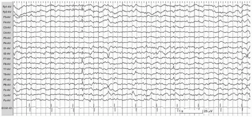
4. What is the finding in the EEG recording shown in Figure 6.1?
A. Bilateral epileptiform spikes
C. Benign sporadic sleep spikes (BSSS)
D. Artifact
View Answer
4. (C): This EEG tracing (Fig. 6.1) is displayed in an average referential montage in light sleep.
There are two low voltage spike-like transients, one predominantly right and the other left temporal. These negative transients exhibit a smaller positivity on the opposite side. These findings are consistent with BSSS also known as small sharp spikes (SSS) or benign epileptiform transients of sleep (BETS).
They are normally seen in approximately 25% of adults during drowsiness and light sleep. They typically have a short duration (<50 ms), low voltage (<50 µV), and a single monophasic or diphasic morphology. They have a broad field maximally seen in the temporal regions, and occasionally have an oblique transverse dipole as in this case. They may have a small aftergoing slow wave.
They are distinguished from epileptiform abnormalities by their morphology, absence of background disturbance, tendency to shift between the two sides, and most importantly by their disappearance during deeper stages of sleep. (Blume, Kaibara and Young 2006, pp. 164, 185; Ebersole and Pedley 2003, p. 241)
5. The EEG of a 52-year-old man reveals an occipital alpha rhythm of 8 Hz. This is considered a normal finding.
A. True
B. False
View Answer
5. (B): The normal frequency range of the occipital alpha rhythm in adults ranges from 8 to 13 Hz. However, the incidence of an 8-Hz occipital alpha rhythm is seen in <1% of the population. This statistically raises the suspicion of a slowed posterior rhythm. Consequently, an occipital rhythm frequency at the low end of the spectrum (such as 8 Hz) is highly suggestive of an EEG abnormality and is commonly referred to as abnormal occipital rhythm. (Ebersole and Pedley 2003, pp. 102-103)
6. Which of the following invasive EEG techniques is not used for lateralization of the epileptogenic zone in patients with bilateral epileptiform discharges?
A. Subdural strips
B. Subdural grids
C. Depth electrodes
D. Foramen ovale electrodes
View Answer
6. (B): All of the above invasive methods except subdural grids can be used for lateralization of ictal foci, especially in suspected mesial temporal lobe epilepsy.
Foramen ovale electrodes, bilateral subtemporal strips, and depth electrodes can be used in determining lateralization of ictal foci when scalp EEG lateralization is inconclusive.
Subdural grids require a craniotomy, which should not be bilateral. Subdural grids are only used for definitive localization of ictal zone but not for lateralization. (Ebersole and Pedley 2003, pp. 642-646)
7. What is a typical ictal onset frequency for seizures presumed to start in the hippocampus?
A. Fast alpha
B. 5 Hz
C. 2 to 4 Hz
D. Generalized attenuation
View Answer
7. (B): Seizures presumed to start in the hippocampus commonly present with ictal onset of 5-Hz frequency involving the sphenoidal or zygomatic electrodes. The rhythmic ictal discharge appears within 30 seconds of the clinical seizure onset. This ictal pattern has a high lateralizing value (>95%) in mesial temporal lobe epilepsies. (Ebersole and Pedley 2003, pp. 562-564)
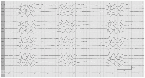
8. The EEG of a 42-year-old comatose male is shown in Figure 6.2. Which of the following is true about this EEG pattern?
A. It is specific for postanoxic brain injury
B. It can be responsive to stimulation
C. It can be seen in drug-induced coma
View Answer
8. (C): This EEG tracing (Fig. 6.2) is displayed in a longitudinal bipolar montage.
There are periodic one-second bursts of synchronous high amplitude theta activity separated by 2 to 3 seconds of complete EEG attenuation. These findings are referred to as burst-suppression pattern. This EEG pattern is nonspecific, most commonly seen in severe brain anoxia, drug-induced coma or severe hypothermia. Burst-suppression pattern is not responsive to stimulation. It has a very poor prognosis when it is a result of anoxic brain injury. Patients with this EEG pattern will be in coma. (Ebersole and Pedley 2003, pp. 355, 408-409; Blume, Kaibara and Young 2002, pp. 487-489)
9. During an absence seizure, generalized 3 Hz spike-and-wave discharge is faster at:
A. Onset
B. Mid-discharge
C. End
D. Does not change with evolution of the ictal discharge
View Answer
9. (A): The generalized 3-Hz spike-and-wave complex associated with absence seizures is faster at the beginning of the ictal discharge. The frequency at onset is approximately 3 to 4 Hz. It slows down to 2.5 to 3 Hz by the end of the discharge. (Wyllie, Gupta and Lachhwani 2005, p. 307)
10. Which of the following best describes the photomyoclonic response?
A. Spike-and-wave activity
B. Muscle artifact
C. Frontal intermittent rhythmic delta activity (FIRDA)
D. Absence of posterior rhythm
View Answer
10. (B): Unlike the photoconvulsive response, the photomyoclonic response is not cerebral. It consists of contraction of frontal scalp muscles causing electromyographic (EMG) activity that is often time-locked to the light stimulus. It has no association with epilepsy. (Abou-Khalil and Misulis 2006, pp. 67-69)
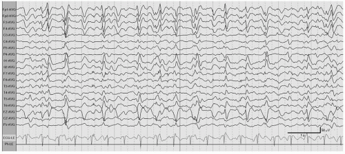
11. The EEG recording shown in Figure 6.3 is most consistent with the diagnosis of:
A. Subdural hemorrhage
B. Hepatic encephalopathy
C. Subacute sclerosing panencephalitis (SSPE)
D. Vertex waves in normal sleep
View Answer
11. (B): This EEG tracing (Fig. 6.3) is displayed in an average referential montage.
There are trains of triphasic waves characterized by a small negative phase and a high voltage positive phase, followed by a lower voltage slower negative phase. Triphasic waves usually predominate in the anterior leads and frequently display an anterior-posterior phase lag of 25 to 140 ms. Triphasic waves are not specific but are commonly seen with toxic-metabolic encephalopathies, notably hepatic. (Ebersole and Pedley 2003, pp. 354-355, 412-415)
12. Temporal spikes are activated during:
A. Photic stimulation
B. Sleep
C. Wakefulness
D. Hyperventilation
View Answer
12. (B): Temporal spikes have a strong association with epilepsy. Sleep, especially non-rapid eye movement (REM) sleep, markedly activates temporal spikes and sharp waves. It is estimated that >90% of patients with temporal lobe seizures will have epileptiform discharges during sleep. (Wyllie, Gupta and Lachhwani 2005, pp. 173-174)
13. All of the following is true about photoparoxysmal response except:
A. Always outlasts photic stimulation
C. Its frequency is independent of the photic frequency
D. Is often associated with generalized epilepsy
View Answer
13. (A): The photoparoxysmal or photoconvulsive response is triggered by flashing lights during photic stimulation. It usually consists of generalized spike-and-wave discharges that may or may not outlast the light stimulation. Its frequency is usually slower than the frequency of the flashing lights. It is most often associated with generalized epilepsy. (Abou-Khalil and Misulis 2006, p. 68)
14. During a presurgical work-up, which of the following tests involve exposure to radiation?
A. Positron emission tomography (PET)
B. Single photon emission tomography (SPECT)
C. Functional magnetic resonance imaging (fMRI)
D. A and B
View Answer
14. (D): SPECT and PET are often performed during epilepsy monitoring for localization of ictal zones in presurgical evaluations.
SPECT is commonly performed with the use of 99 mTechnetiumhexamethylpropylene amine oxime (99 mTc-HMPAO) tracer, a nuclear isomer that emits gamma rays.
PET uses metabolically active molecules that emit positrons, which rapidly get annihilated upon meeting electrons, emitting gamma rays. The molecule most commonly used clinically for this purpose is 18F-fluorodeoxyglucose (FDG). (Wyllie, Gupta and Lachhwani 2005, pp. 993-1008)
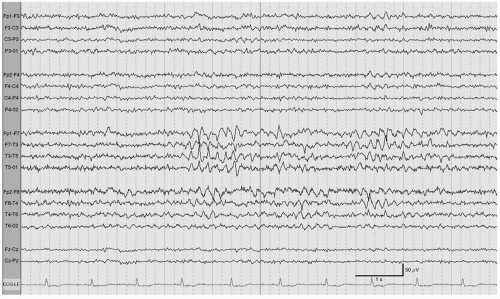
15. The EEG recording shown in Figure 6.4 is that of a 40-year-old man during drowsiness. The EEG finding is most consistent with:
A. Partial seizure
B. Trains of temporal sharp waves
C. Normal variant
D. Temporal intermittent rhythmic delta activity (TIRDA)
View Answer
15. (C): This EEG tracing (Fig. 6.4) is displayed in a longitudinal bipolar montage.
There are bursts of rhythmic theta activity with notched appearance during drowsiness. This activity is predominant in the left mid-temporal region. This EEG pattern is known as rhythmic midtemporal discharges (RMTD) or rhythmic temporal theta burst of drowsiness (RTTBD). RMTD was also previously referred to as psychomotor variant. This EEG pattern can be bilateral or independent over the two hemispheres as in this case. It is a normal variant of drowsiness in adolescents and adults. Unlike ictal discharges, this EEG pattern does not interrupt the background and lacks evolution in frequency, amplitude, or distribution, which is typical of ictal events. (Ebersole and Pedley 2003, pp. 236-238)
16. During intraoperative electrocorticography, which agent can be used to activate epileptiform discharges?
A. Pentobarbital
B. Methohexital
C. Sevoflurane
D. Propofol
View Answer
16. (B): Electrocorticography is sometimes performed intraoperatively during epilepsy surgery. It records brain activity directly from the cortex. Recording of epileptiform discharges may help to delineate margins of the epileptogenic zone for resection or placement of subdural electrodes. In the absence of spontaneous epileptiform discharges, methohexital (a rapid short acting barbiturate) can be used to activate epileptiform discharges. This effect occurs only at low doses of methohexital; higher doses of this drug will suppress brain activity, similarly to other anesthetic drugs. (Ebersole and Pedley 2003, pp. 267, 686-689)
17. Hypnic jerks are associated with:
A. Spike-and-wave discharges
B. K-complexes
C. Photic stimulation
View Answer
17. (B): Myoclonic or hypnic jerks occur during the transition from drowsiness to sleep in normal patients. It is often accompanied by vivid sensory feelings like falling off the bed. Hypnic jerks are associated with vertex waves and K-complexes.
When they become frequent, they can disturb sleep and are then classified as periodic limb movement disorders. (Daube 2002, pp. 407-408)
18. Lambda waves are seen during:
A. Heightened alertness
B. Drowsiness
C. Visual scanning
D. Rapid eye movement (REM) sleep
View Answer
18. (C): Lambda waves are positive occipital notched waves seen during visual scanning of a complex picture. Saccadic eye movements are usually involved in the generation of these waves. Lambda waves are attenuated with eye closure. They can be asymmetrical, leading to misinterpretation as an abnormality.
Lambda waves share the same morphology as POSTS, which are exclusively seen during sleep. (Ebersole and Pedley 2003, p. 126; Blume, Kaibara and Young 2002, pp. 88-90)
19. All of the following is true about EEG changes during a syncopal episode except:
Stay updated, free articles. Join our Telegram channel

Full access? Get Clinical Tree


