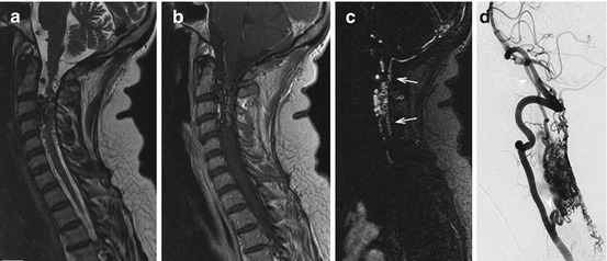
Fig. 5.1
An example of high-resolution CE-MRA in a patient with a spinal dural arteriovenous fistula (SDAVF, more details are given in Chap. 19): a centromedullary edema with faint enhancement and perimedullary dilated vessels are shown on conventional T2-weighted (a) and fat-saturated T1-weighted images (b). MIP reconstructions of the first-pass CE- MRA images clearly depict the early venous filling (c, arrows) confirming the shunt of the SDAVF. The level of the radiculomeningeal artery supplying the shunt is also demonstrated (arrow head) and was later proven by selective X-ray angiography (d) (Courtesy of Timo Krings, Professor of Radiology, Diagnostic and Interventional Neuroradiology, Toronto Western Hospital & University Health Network)

Fig. 5.2
An example of first-pass CE-MRA in a patient with a spinal arteriovenous malformation (SAVM), which appears as a series of multiple flow voids in and around the cervical cord on a T2-weighted image (a) with irregular enhancement on a contrast-enhanced T1-weighted image (b). First-pass CE-MRA clearly demonstrates the nidus with a plexiform network of arteries and draining veins extending into the subarachnoid space (c, arrows). The SAVM was confirmed by selective angiography (d) which also shows the supplying dilated anterior spinal artery and additional segmental supply from the vertebral artery (arrow heads) (Courtesy of Timo Krings, Professor of Radiology, Diagnostic and Interventional Neuroradiology, Toronto Western Hospital & University Health Network)
Even though X-ray based catheter angiography (digital subtraction angiography, DSA) is the gold standard for diagnosing and classifying spinal vascular lesions, information on spinal cord vascular pathology obtained from a preceding CE-MRA scan can facilitate DSA examination. Specifically, knowledge of the feeding arteries, which may originate from the costocervical or thyrocervical trunks, vertebral or any intercostal lumbar and iliolumbar artery, may not only shorten time of intervention but also reduce the risk of complications and radiation exposure. However, a single spinal MRA examination can only cover a limited part of the spine, as the craniocaudal extension of the field of view (FOV) should be small enough to allow high-resolution angiograms.
5.2 Diffusion Imaging
Diffusion is the random motion of molecules through a medium. The freedom water molecules have to diffuse depends on their local environment. For example, water within cells is much more restricted than extracellular water. Since diffusion characteristics can change with disease, it is useful to be able to probe this phenomenon. In MRI the signal can be made sensitive to diffusion by the addition of large gradient pulses after the RF excitation pulse, which first encode and then decode the spatial position of the water molecules a short time later. The spatial position of water molecules that diffuse rapidly along the direction of the applied gradient is not properly decoded, leading to dephasing and thus signal loss. Conversely, water molecules that are in restricted environments cannot diffuse very far and therefore the signal strength is not greatly affected by the diffusion gradients. Comparison of images with and without diffusion weighting allows the calculation of an apparent diffusion coefficient (ADC) in each voxel. A high ADC corresponds to larger net displacement of the water molecules (i.e. relatively unrestricted) and therefore low signal strength in the diffusion-weighted image.
Stay updated, free articles. Join our Telegram channel

Full access? Get Clinical Tree


