Anatomy and physiology
The nervous system serves as the body’s communication network. It processes information from the outside world, through the sensory portion, and coordinates and organizes the functions of all other body systems. Its far-reaching effects can be seen when patients, who suffer from diseases of other body systems, develop neurologic impairments related to the disease. For example, the patient who has heart surgery could suffer a stroke.
The neurologic system is divided into the central nervous system (CNS), the peripheral nervous system, and the autonomic nervous system (ANS). Through complex and coordinated interactions, these three systems integrate all physical, intellectual, and emotional activities. Understanding how each works is essential to conducting an accurate neurologic assessment.
NERVOUS SYSTEM CELLS
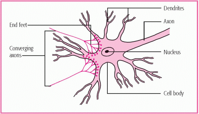
Two major cell types, neurons and neuroglia, compose the nervous system. The neuron is the fundamental unit of the nervous system. It’s a highly specialized conductor cell that transmits and receives electrochemical nerve impulses. Delicate, threadlike nerve fibers extend from the central cell body to transmit impulses—axons carry these impulses away from the cell body, whereas dendrites carry impulses to it. Most
neurons have multiple dendrites but only one axon. (See Structure of the neuron.)
neurons have multiple dendrites but only one axon. (See Structure of the neuron.)
Sensory (afferent) neurons transmit impulses from special receptors to regulate activity in the brain and spinal cord. Motor (efferent) neurons transmit impulses from the CNS to regulate activity in muscles or glands, whereas interneurons (connecting or association neurons) shuttle signals through complex pathways between sensory and motor neurons. Interneurons account for 99% of all the neurons in the nervous system and include most of the neurons in the brain itself.
Neuroglial cells, or glial cells (derived from the Greek word for glue because they hold the neurons together), serve as the supportive cells of the CNS and form roughly 40% of the brain’s bulk. The four types of neuroglial cells include:
Astroglia, or astrocytes, exist throughout the nervous system and form part of the blood-brain barrier. They supply nutrients to the neurons and help maintain their electrical potential.
Ependymal cells line the brain’s four ventricles and the choroid plexus and help produce cerebrospinal fluid (CSF).
Microglia phagocytize waste products from injured neurons and are deployed throughout the nervous system.
Oligodendroglia support and electrically insulate CNS axons by forming protective myelin sheaths.
CENTRAL NERVOUS SYSTEM
The CNS includes the brain and the spinal cord. These two major structures collect and interpret voluntary and involuntary motor and sensory stimuli. (See Major structures of the central nervous system, page 4.)
The brain and spinal cord engage in an intricate network of interlocking receptors and transmitters, ultimately forming a dynamic control system—a “living computer”—that oversees and regulates every mental and physical function. Indeed, from birth to death, the CNS efficiently organizes the body’s affairs —controlling the smallest action, thought, or feeling; monitoring communication and the instinct for survival; and allowing introspection, wonder, and abstract thought.
The brain, the center of the CNS, is a large, soft mass of nervous tissue that’s housed within the cranium and supported by the meninges. The brain and spinal cord are protected by bone (the skull and vertebrae), which cushions CSF, and three membranes:
dura mater, or outer sheath, made of tough, white fibrous tissue
arachnoid membrane, the delicate, lacelike middle layer
pia mater, the inner meningeal layer, consisting of fine blood vessels held together by connective tissue. (This membrane is thin and transparent and clings to the brain and spinal cord surfaces, carrying branches of the cerebral arteries deep into the brain’s fissures and sulci.)
Between the dura matter and the arachnoid membrane is the subdural space; between the arachnoid membrane and the
pia mater is the subarachnoid space. Within the subarachnoid space and the brain’s four ventricles is the aforementioned CSF—a substance comprised of water and traces of organic materials (especially protein), glucose, and minerals. CSF is formed from blood in capillary networks called choroid plexi, which are located primarily in the brain’s lateral ventricles.
CSF is eventually reabsorbed into the venous blood through the arachnoid villi, in dural sinuses on the brain’s surface.
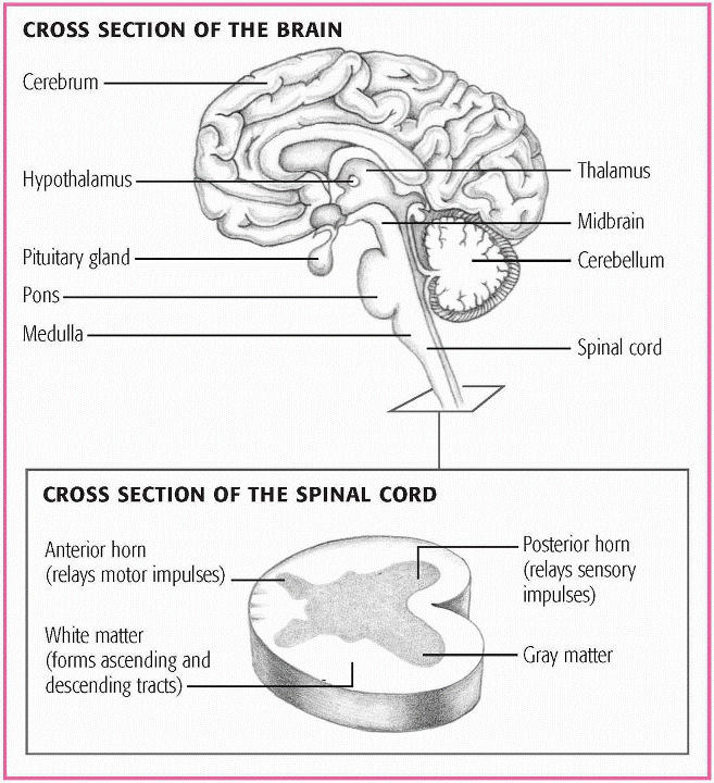
MAJOR STRUCTURES OF THE CENTRAL NERVOUS SYSTEM
This illustration shows a cross section of the major structures of the central nervous system—the brain and spinal cord. The brain joins the spinal cord at the base of the skull and ends between the first and second lumbar vertebrae. Note the H-shaped mass of gray matter in the spinal cord.
|
Brain
The brain consists of the cerebrum, or cerebral cortex, the brain stem, the cerebellum, the limbic system, and the reticular activating system (RAS). It collects, integrates, and interprets all stimuli and initiates and monitors voluntary and involuntary motor activity.
CEREBRUM
The cerebrum, the largest portion of the brain, houses the nerve center that controls motor and sensory functions and intelligence. It’s encased by the skull and enclosed by three membrane layers called meninges. If blood or fluid accumulates between these layers, pressure builds inside the skull and compromises brain function. The surface layer of the cerebrum is the cerebral cortex, which is composed of unmyelinated cell bodies called gray matter. Additionally, the cerebrum has a rolling surface made up of convolutions, called gyri, and creases or fissures, called sulci.
The cerebrum consists of a left and right hemisphere, joined by the corpus callosum—a mass of nerve fibers that allows for communication between the hemispheres and for sharing learning and intellect. However, these two hemispheres don’t share equally; one always dominates, giving one side control over the other. Because motor impulses descending from the brain through the pyramidal tract intersect in the medulla, the right hemisphere controls the left side of the body, whereas the left hemisphere controls the right side of the body.
Several fissures divide the cerebrum into lobes, each of which is associated with specific functions. To that end, each hemisphere is divided into four lobes, based on anatomic landmarks and functional differences. The lobes are named for the cranial bones that lie over them: the frontal, temporal, parietal, and occipital.
The frontal lobe influences personality, judgment, abstract reasoning, social behavior, language expression, and movement.
The temporal lobe controls hearing, language comprehension, and the storage and recall of memories, although some memories are stored throughout the brain.
The parietal lobe interprets and integrates sensations, including pain, temperature, and touch. It also interprets size, shape, distance, and texture. The parietal lobe of the non-dominant hemisphere, usually the right, is especially important for awareness of body schema or shape.
The occipital lobe functions primarily in interpreting visual stimuli.
In addition, cranial nerves (CNs) I and II originate in the cerebrum. The cerebrum is considered the area involving upper motor neuron function. (See The cerebrum and its functions.)
The diencephalon, a division of the cerebrum, contains the thalamus and hypothalamus. The thalamus is a relay station for sensory impulses as they ascend to the cerebral cortex. Its functions include primitive awareness of pain, screening of incoming stimuli, and focusing of attention and emotional response. The hypothalamus lies beneath the thalamus and is an autonomic center that has connections with the brain, spinal cord, ANS, and pituitary gland. It regulates temperature, appetite, blood pressure, breathing, sleep patterns, and peripheral nerve discharges that occur with behavioral and emotional expression. It also partially controls pituitary gland secretion and stress reaction.
BRAIN STEM
The brain stem lies below the diencephalon and is divided into the midbrain, pons, and medulla. These three parts provide two-way conduction between the spinal cord and brain.
The brain stem relays messages between the parts of the nerous system. It has three main functions that include producing the rigid autonomic behaviors necessary for survival,
such as increasing the heart rate and stimulating the adrenal medulla to produce epinephrine; providing the pathways for nerve fibers between higher and lower neural centers; and serving as the origin for 10 of the 12 pairs of cranial nerves.
such as increasing the heart rate and stimulating the adrenal medulla to produce epinephrine; providing the pathways for nerve fibers between higher and lower neural centers; and serving as the origin for 10 of the 12 pairs of cranial nerves.
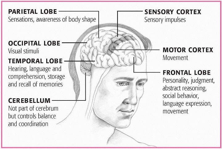
THE CEREBRUM AND ITS FUNCTIONS
The cerebrum is divided into four lobes, based on anatomic landmarks and functional differences. The lobes – parietal, occipital, temporal, and frontal – are named for the cranial bones that lie over them.
This illustration shows the locations of the cerebral lobes and explains their functions. It also shows the location of the cerebellum.
|
Stay updated, free articles. Join our Telegram channel

Full access? Get Clinical Tree


