Figs. 1.1, 1.2, and 1.3
Sixth cervical vertebra is characterized for the anterior tubercle of the transverse process which is more developed and prominent (Chassaignac tubercle): landmark for the common carotid (ligature), for the inferior tiroid artery and for the vertebral artery
Cervical Spine Joints
Joints between vertebrae are made for making spine mobile.
Vertebrae which make up the different part of the spine have joints that allow to the different parts of spine different movements.
Two sections of cervical spine, “superior cervical spine ”, which includes occipital bone, atlas and axis and the “inferior cervical spine ” which extendes from the inferior edge of the axis to the superior edge of the first thoracic vertebra, have different joints that functionally complete each others allowing movements like rotation, inclination, flexion, extension of the head.
The occipito-atlas joints realize flexion-extension movements on sagittal plane and inclination on frontal plane. They involve occipital bone condyles and the concave articular facets located on lateral masses of the atlas. These joints, defined condylartrosis, have two axis of movement and two grades of freedom. This joint is made of two bones and a fibrous capsule , covered by a synovial membrane, which insert in the boundary of the occipital condyles and on the border of the superior articular facet of the atlas. The articular capsule medially is thin and lateral is thicker and it is enforced by the anterior atlas-occipital membrane which extends from the posterior edge of the foramen magnum to the superior edge of the posterior arch of the atlas.
The anterior atlas-occipital membrane has a fibrous band where origins the anterior longitudinal ligament and the posterior occipital membrane is crossed by the first cervical nerve and by the vertebral artery.
Between the occipital condyle and the atlas the first pair of spinal nerves emerge and they will form the cervical plexus.
This joint alone realizes almost the 50 % of the flexion and extension of the head involving for the rest the whole cervical section.
The atlas-axis joint is a diartrosis with no inter vertebral disk between C1 and C2. It’s formed by the atlas, axis lateral joints atlas-axis medial joint and atlas-odontoid.
The lateral atlas-axis joint is a diartrosis of artrodia type and it’s made by the inferior articular facets of the lateral masses which are slightly concave and by the superior articolar facets of the axis which are slightly convesse. The articular capsule is inserted on the lateral edges of the articular cartilagines and it’s enforced anteriorly by the anterior longitudinal ligament and posteriorly by the yellow ligaments and postero medially by the accessorius ligament which arises from the posterior section of the body of the axis and insert on the posterior face of the lateral masses of the atlas.
The medial atal-axis joint is a pivot joint between the posterior facet of the anterior arch of the atlas covered by articular cartilage ad the articular facet of the odontoid covered also by articular cartilage.
The joint is stabilized posteriorly by a fibrous lamina, the “transverse ligament ” which surrounds the tooth.
And extendes between the two lateral masses of the atlas realizing in this way a osteo-fibrous ring made anteriorly by the anterior arch of the atlas and posteriorly by the transverse ligament.
From the medial section of the ligament start some fibers that go up to insert on the basilar part of the occipital bone, superior longitudinal ligament , and down on the posterior face of the axis, inferior longitudinal ligament (Fig. 1.4) [9, 10]
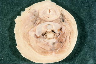
Fig. 1.4
From the medial section of the ligament start some fibers that go up to insert on the basilar part of the occipital bone, superior longitudinal ligament, and down on the posterior face of the axis, inferior longitudinal ligament
The articulation of the “inferior cervical spine” are artrodial joints and between the vertebral bodies intervertebral disks are present.
This section is specialized in flexion -extension movements and lateral flexion : lateral flexion is made by c3-c5 joint while flexion-extension by the C4-C6.
In the inferior cervical spine the joints have common features to the whole spine except sacrum. There are inter-somatic junctions between vertebral bodies esured by the presence of the intervertebral disk and the zigoapofisary joints between the superior articular processes of a vertebra and the inferior articular processes of the lower vertebra. The intersomatic joints are sinartrosis of sinfisys type which are made between the surface of the body of a vertebra and the inferior one of the body of the upper vertebra covered by ialine cartilage.
The shape of the disks is like the one of the vertebral bodies whose is in between and contribute on the flexion-extension movement and lateral flexion of this section and to form the cervical lordosis so the diskes are thicker anteriorly. Discs and vertebral bodies are united and stabilized by the anterior and posterior longitudinal ligaments.
The anterior longitudinal ligament is a fibrous tape which arises from the occipital bone and from th body of the axis and running in the inside of the vertebral channel, it inserts on the posterior surface of the vertebral bodies.
The intervertebral disk is made by a central part called nucleus polposus and by a peripheral part called annulus fibrous. Nucleus polposus is made by a deformable and incompressible gel made by mucopolysaccharides and water. Hydrophilic properties of proteoglycans depend on quantity and quality of mucopolysaccharides.
Idrophily of nucleus polposus determines vertebral resistance to mechanical loading whilst fibro-cartilaginous ring which it’s made by anular fibres, allows flexion movements.
These structural features of disk are very important especially following spinal trauma because herniae of nucleus polposus into the specus vertebralis may happen with compression of nerve roots.
Zigoapofisary joints are artrodiae that allow movements of slipping between superior and inferior vertebral processes of two close vertebrae. Superior articular processes, covered by ialine cartilage, run up and backward whilst inferior one run down and forward and are covered by a thin articular capsule that ends on the edge of articular cartilage.
We distinguish:
Intrinsic Muscles of the Cervical Spine, in which muscles have their attachment only on the vertebrae:
Interspinous muscles
Intertrasversarii cervici muscles
Multifidus muscle
Short and long rotator muscles
Semispinalis Capitis muscle
Spinalis cervicis muscle
Longissimus Cervicis muscle
Longus cervicis muscle
Inferior obliquus cervicis muscle
Extrinsic muscles , in which muscles have their insertion both in cervical spine and in other skeletal segments:
Spinalis capitis muscle
Semispinalis capitis muscle
Longissimus capitis muscle
Iliocostalis muscle
Rectus capitis major and minor muscles
Obliquus capitis superior muscle
Rectus capitis anterior and lateral muscle
Levator scapulae muscle
Splenius capitis and cervicis muscles
Serratus posterior superior muscle
Rhomboideus minor muscle
Trapezius muscle
Neck proprii muscles –> (excluding cervical spine):
Suprahyoid muscles: digastric, stylohyoid, mylohyoid and geniohyoid
Infrahyoid muscles: omohyoid, sternocleidohyoid, sternothyroid and sternohyoid muscles
Sternocleidomastoid muscleRelated posts:
 Pathophysiology of Cervical Pain: Evolution and Treatment
 Anterior Cervical Approach: Decompression and Fusion with Cages
 Cervical PLDD (Percutaneous Laser Discectomy). Ten Years Experience
 Anterior Cervical Decompression and Fusion with Autologous Bone Graft
 Odontoid Screw Fixation
 Injuries of the Middle and Lower Cervical Spine

Stay updated, free articles. Join our Telegram channel

Full access? Get Clinical Tree


