, Nima Etminan1 and Daniel Hänggi1, 2
(1)
Neurochirurgische Klinik, Universitätsklinikum Düsseldorf, Düsseldorf, Germany
(2)
Medical Art Christine Opfermann-Rüngeler, Zentrum für Anatomie Heinrich Heine Universität, Düsseldorf, Germany
7.1 Aneurysms of the Middle Cerebral Artery Main Bifurcation
7.1.1 General Considerations
Middle cerebral artery (MCA) aneurysms are fairly peripheral to the arterial tree, occurring close to the cranial surface. They are often broad based and rarely lead to a hematoma in the temporal lobe or Sylvian fissure. (Compare also Sect. 7.2.) These factors are in favor of the microsurgical approach, although many MCA aneurysms can technically also be controlled with modern endovascular systems [1, 2].
The location and projection of the aneurysms is important for approach planning. As outlined earlier, we prefer a tailored transsylvian approach in which the M2 segments are identified first and then followed backward to the bifurcation and the neck of the aneurysm.
Planning the approach carefully is important for safe and efficient control of MCA aneurysms. It is important to have a mental concept of the location of the fundus in relation to the bifurcation. The most frequent projections are downward (type 1), with the dome being buried in the temporal lobe (Fig. 7.1), or lateral (type 2), with the aneurysm being more or less a lateral extension of the M1 segment (Fig. 7.2). These latter aneurysms lie in the Sylvian fissure. Type 3 aneurysms lie within the plane of the M2 trunks (Fig. 7.3). The rare, upward-directed type 4 aneurysms are buried in the frontal lobe (Fig. 7.4). The approach should plan to avoid the dome of the aneurysm. Therefore, for the frequent projections of types 1 and 2, the initial dissection of the Sylvian fissure should follow the frontal operculum down to the superior trunk of the MCA. The position of the dome also must be kept in mind when using retractor blades on the temporal operculum.
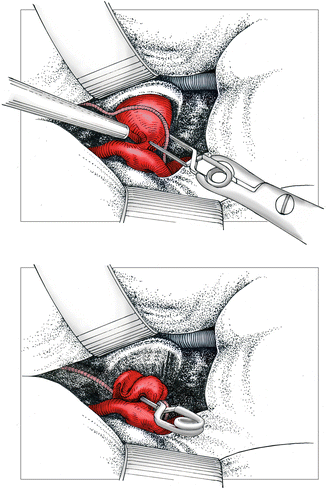
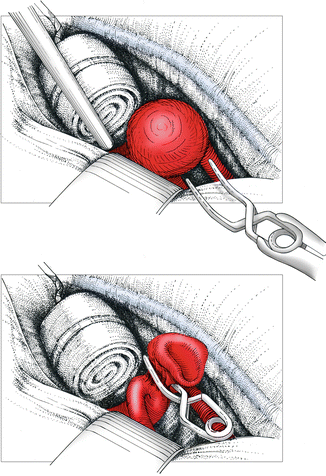
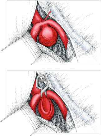
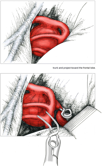
Fig. 7.1
Left-sided type 1 middle cerebral artery (MCA) aneurysm with the dome projecting from the bifurcation toward the temporal lobe. Note the anterior temporal artery curving around the aneurysm. Venous drainage is somewhat atypical in this case in that the main vein runs within the Sylvian fissure. Final clip position following separation of the anterior temporal artery from the aneurysm neck. The plane of the clip is approximately parallel to the plane of the bifurcation
Fig. 7.2
Right-sided type 2 MCA aneurysm. The aneurysm is essentially an elongation of the afferent M1 segment and lies within the Sylvian fissure. A rolled cotton pad is used to hold the Sylvian fissure open and to minimize retractor pressure. Final clip position with the clip blades in the plane of the M2 trunks
Fig. 7.3
Right-sided type 3 MCA aneurysm. The dome lies between the M2 trunks. Final clip position with the clip perpendicular to the plane of the M2 trunks
Fig. 7.4
Left-sided type 4 MCA aneurysm. The dome is hidden behind the superior trunk and projects toward the frontal lobe. Following temporary clipping of M1, a curved clip is directed around the superior trunk
7.1.2 Dissection and Clipping
After opening the dura, the Sylvian fissure is subsequently split on the frontal side of the Sylvian vein. One to three venous branches crossing the fissure usually must be coagulated and divided. It is important to split the Sylvian fissure all the way down to the sphenoid ridge to allow for separation of the frontal and temporal lobes. Sufficient dissection at this stage avoids the later necessity to use excessive retraction on the frontal operculum. The superior trunk of the MCA is then identified in the depth of the Sylvian fissure. This artery is then followed proximally to the MCA bifurcation.
To secure proximal control, it is important to keep in mind the course of M1 and the projection of the aneurysm. Most MCA aneurysms are directed more or less toward the temporal lobe. A minority are attached to the frontal side. Therefore proximal control usually is achieved by dissection of the MCA bifurcation along the superior trunk. M1 is identified between the main branches in the case of a high-running M1 segment or in front of the superior trunk in the case of a low-running M1.
Whenever possible, we prefer not to use any brain retractors at all for MCA aneurysms, but sometimes the brain is so swollen that it is impossible to gain sufficient working space without a brain retractor on the frontal operculum. The temporal operculum frequently covers a temporally projecting aneurysm, and it is sometimes necessary to use a small spatula on the temporal operculum during the clipping period.
Temporary clipping of M1 should be used generously whenever the neck of the aneurysm is wider than the diameter of M1. When applying the temporary clip, the origin of the lateral lenticulostriate arteries must be spared.
The final position of the aneurysm clip will be in front of the MCA bifurcation for type 1 projections of the dome and above the bifurcation for type 2. The clip will lie in a plane more or less parallel to the plane of the bifurcation (Fig. 7.1). Dissection of the aneurysm neck is relatively easy with these projections, as the neck is not in direct contact with the M2 trunks.
With type 1 and type 2 aneurysms, however, the anterior temporal artery requires attention. It usually originates on the distal M1 segment and then curves around the aneurysm neck or dome. It must be separated from the neck and spared during clipping. Occlusion of the anterior temporal artery usually leads to infarction of the lateral anterior temporal lobe.
On the other hand, type 3 aneurysms lie between the M2 trunks and need to be separated from these trunks prior to clip application. Here the final clip position will be in a plane more or less perpendicular to the plane of the bifurcation (Fig. 7.3).
The rare upward-projecting aneurysms (type 4) require dissection on the frontal side of the superior MCA main trunk. The lateral lenticulostriate arteries often originate close to the neck of these aneurysms, and particular attention is necessary during dissection and clipping (Fig. 7.5).
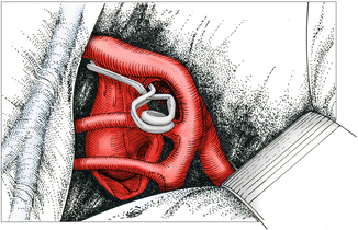
Fig. 7.5


Final clip position for type 4 with the clip parallel to the plane of the M2 trunks. Particular attention must be paid to the lenticulostriate arteries, which are often close to the neck of type 4 aneurysms (not shown)
Related posts:

Stay updated, free articles. Join our Telegram channel
Full access? Get Clinical Tree
