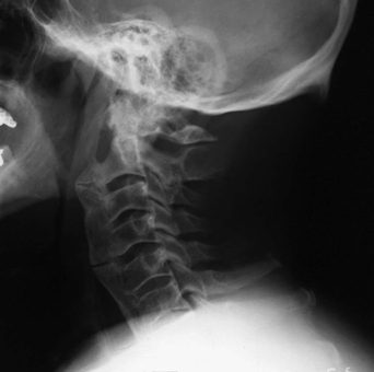
77 This 69-year-old man fell forward 6 months ago and began complaining of neck pain. He remains neurologically intact and without pain. C-spine lateral x-ray demonstrates diffuse ankylosing hyperostosis with ossifications involving the anterior aspect of the spine. The osteophytic bridge is disrupted at C2-C3 and C4-C5 (Fig. 77-1). The patient does not move on flexion extension films, and his alignment is intact. Diffuse idiopathic skeletal hyperostosis (DISH) Because there is no translational instability, subluxation, cord signal changes on magnetic resonance imaging (MRI), or other evidence of misalignment, and the patient is pain free, his collar was discontinued. His spine is stable after a fall 6 months ago. DISH is an ossifying disease of unknown etiology that affects ~10% of patients in the sixth decade of life. It is characterized by “flowing” calcifications and “claw” spurring of the anterolateral aspects of the involved vertebrae. DISH tends to affect the lumbar spine, usually at the L4 level. End-plate osteophytes and disc collapse are seen, and patients report low back pain. The cervical spine is also affected and is often an incidental finding or thought to be the etiology of dysphagia, and less often, myelopathy. Differentiating DISH of the lumbar spine from degenerative stenosis can be nuanced. Leroux et al (1992) have established the following criteria as indicative for DISH: anterior or posterior lateral margin osseous proliferation, proliferation of the nonarticular aspects of the posterior apophysis, and ossification of the posterior articular capsule and posterior ligaments.
Ankylosing Hyperostosis of Forestier and Rotes-Querol
Presentation
Radiologic Findings
Diagnosis
Treatment
Discussion

Ankylosing Hyperostosis of Forestier and Rotes-Querol
Only gold members can continue reading. Log In or Register to continue

Full access? Get Clinical Tree


