(1)
Cedars-Sinai Spine Center, Los Angeles, CA, USA
Anterior exposure of the cervicothoracic spine (C7 to T2) presents a significant challenge because of the limited access afforded by anatomical considerations. The junction of the cervical and thoracic spine not only represents the apex of two physiologic curvatures but is also crowded by important neurovascular structures, the trachea and the esophagus at the narrowed thoracic outlet. Median sternotomy as well as partial resection of the manubrium and part of the clavicle on one side have been described but may not be necessary for adequate exposure because of recent improvements in instrumentation and technique. In addition, the increasing familiarity of many surgeons with the anatomy of the area makes less radical approaches more desirable. We present a stepwise approach to the area that may obviate resection of the clavicle or the manubrium, depending on how much exposure can be obtained before proceeding with those steps.
Get Clinical Tree app for offline access

1.
Under general endotracheal anesthesia, the patient is placed supine with a bolster between the scapulae with the head turned to the right and the neck slightly extended. A slight reverse Trendelenburg position may help reduce venous distension. The vocal cords should be evaluated by the anesthesiologist at the time of intubation to confirm their integrity, and a nasogastric tube is inserted to help localize the esophagus.
2.
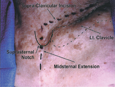
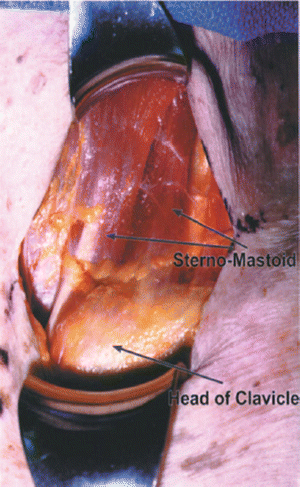
An inverted L-shaped incision is marked off on the skin with the transverse limb 1 to 2 cm above the left clavicle, extending from the lateral border of the sternomastoid muscle to the midline just above the suprasternal notch. From there the vertical limb is marked in the midline extending to the bottom of the manubrium (Fig. 13.1). The skin is initially incised only along the transverse plane, with the vertical portion reserved for use only if exposure is not adequate without resection of the clavicle and part of the manubrium. Subplatysmal flaps are then elevated. These flaps should be wide enough to expose the lower one-third of the sternomastoid and strap muscles superiorly and extend inferiorly 3 to 4 cm below the clavicle (Fig. 13.2).


Fig. 13.1
The skin incision extension has been marked off with anatomical relationships highlighted. Incision extends from just lateral to the sternomastoid to the midline about 1 to 2 cm above the clavicle

Fig. 13.2
Skin flaps have been elevated superiorly and inferiorly, exposing the distal one-third of the sternomastoid and the distal end of the clavicle
3.
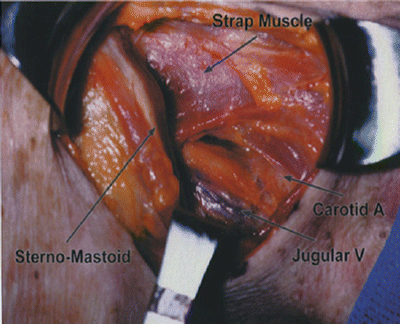
Dissection between the medial border of the sternomastoid and the strap muscles is carried superiorly to the extent of the flap exposure and inferiorly to the insertion at the clavicle. Blunt dissection is then used to create a plane posterior to the sternoclavicular joint to expose the superior mediastinal fat pad. The dissection then mobilizes the sternomastoid muscle with the jugular vein laterally and the strap muscles medially to expose the avascular plane between the carotid sheath laterally and the trachea and esophagus medially (Fig. 13.3). This plane is then developed down to the prevertebral fascia. Very careful dissection in this area is necessary to possibly identify the recurrent laryngeal nerve, which lies in the tracheoesophageal groove.


