(1)
Marina Spine Center, Marina del Rey, CA, USA
The significant anatomical landmarks for differentiating the approaches to the cervical spine presented in Table 1.1 are the sternocleidomastoid muscle, the carotid sheath, and the longus coli muscle (Fig. 1.1) [1 – 8]. Categorization is based on the direction of approach relative to these specific structures, as demonstrated in the table. For example, approach no. 1 is medial to the sternocleidomastoid muscle (therefore, retracting it laterally) and medial to the carotid sheath (therefore, retracting it laterally as well). Approach no. 7 is directed lateral to the sternocleidomastoid muscle and lateral to the carotid sheath. A significant aspect of these approaches is whether to approach the carotid sheath medially or laterally. Approaching the carotid sheath medially and retracting laterally often requires sacrifice of vessels coursing from the carotid sheath to the medial musculovisceral column. Nerves running from lateral to medial also must be retracted. Approaching the carotid sheath laterally and retracting it medially, as in the anterolateral approaches [1 – 5] produces a more avascular plane but may also result in a more limited exposure. Both the anteromedial and the anterolateral approaches have common points of dissection and anatomy.

Table 1.1
Approaches to the Cervical Spine
Sternocleidomastoid Muscle | Carotid Sheath | Longus Coli | ||||
|---|---|---|---|---|---|---|
Medial | Lateral | Medial | Lateral | Medial Lateral | ||
X | X | X | ||||
2. Anterior medial | X | X | X | |||
3. Supraclavicular approach [4] | X | X | X | |||
4. Anterolateral approach to | X | X | X | |||
5. Anterolateral approach to | X | X | X | |||
midcervical spine [2] | ||||||
6. Lateral approach to | X | X | X | |||
midcervical spine [10] | ||||||
7. Lateral approach to | X | X | X | |||
midcervical spine1 | ||||||

Fig. 1.1
The numerous soft tissue structures of the anterior neck. Note the relatively avascular area between the superior thyroid artery and the inferior thyroid artery. The standard approach to the anterior aspect of the spine is lateral to the thyroid gland and medial to the carotid artery
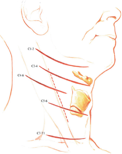
The skin incision must be cosmetically acceptable but efficient (Fig. 1.2). Superficial landmarks used to place the incision over the appropriate level of the spine are (1) C3–C4, which is 1 cm above the thyroid cartilage; and (2) C5–C6, which is at the cricoid cartilage [6]. Other superficial landmarks to be identified are the angle of the jaw, the sternocleidomastoid muscle, the hyoid bone, the cricoid cartilage, the superior border of the thyroid cartilage, and the insertion to the sternocleidomastoid to the clavicle. For best cosmesis, make a 3-cm transverse incision in a skin crease. A longer transverse incision from midline to anterior border of the sternocleidomastoid muscle will allow adequate exposure for three vertebral bodies and two disc levels. The exact pathology and the technical demands of the operation determine the size of the exposure and the structures that must be transected rather than retracted.


