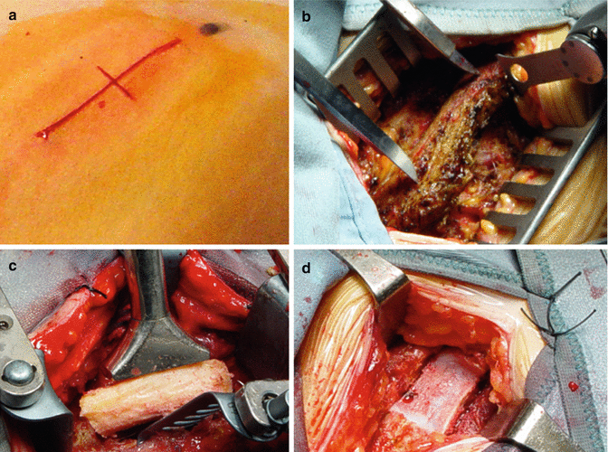
Fig. 9.1
Incision and soft tissue dissection. (a) A transverse skin incision extending from the midline to the medial border of the sternocleidomostoid muscle is adequate to expose up to two intervertebral discs or one vertebral body. An oblique incision along the anterior border of the sternocleidomastoid muscle is used in more extensive procedures. (b) After incision of the platysma and subplatysma dissection, the superficial layer of the deep cervical fascia is entered with exposure of the sternocleidomastoid muscle. Dissection of the middle layer allows exposure of the omohyoid muscle. (c) The carotid artery can be palpated posterior to the sternocleidomastoid muscle. The alar fascia (asterisk) covers the longus colli muscles and separates the vertebral bodies from the trachea and esophagus. (d) The longus colli muscles are clearly exposed and help to identify the midline. (e) A right-angle bent needle is inserted in the disc and a lateral x-ray is obtained to confirm the level. (f) The longus colli muscles are dissected and the retractor blades are inserted. The anterior longitudinal ligament and the anulus fibrosus are incised and removed. cca common carotid artery, lcm longus colli muscle, om omohyoid muscle, scmm sternocleidomastoid muscle
Smith-Robinson Technique
The anterior longitudinal ligament and the anulus fibrosus are incised flush from the edges of the vertebral bodies with a No. 11 scalpel (Fig. 9.2). The anterior osteophytes, when present, are removed with a curved osteotome or rongeurs. The anterior longitudinal ligament lying on opposing vertebral bodies is removed to clearly expose the bony surface. Interbody distraction is obtained by placing distracting pins into the midportion of the vertebral bodies above and below the interspace to be treated. After distraction, the disc and the cartilaginous plates are progressively removed with curette. High-speed drill and Kerrison pounch are used to complete the discectomy and to remove the posterior osteophytes under microscopic view. The posterior longitudinal ligament (PLL ) is generally opened widely to expose the dura and to verify complete removal of disc herniation and optimal resection of osteophytes . We use a nerve hook to open laterally the PLL, which is elevated from the dura and progressively resected with kerrison pouch. When foraminal stenosis is present, the medial aspect of the uncovertebral joint is resected using a No. 1 or 2 Kerrison punch. Bleeding from epidural veins in the neural foramen is controlled with small pieces of surgicel and gentle pressure with cottonoid.
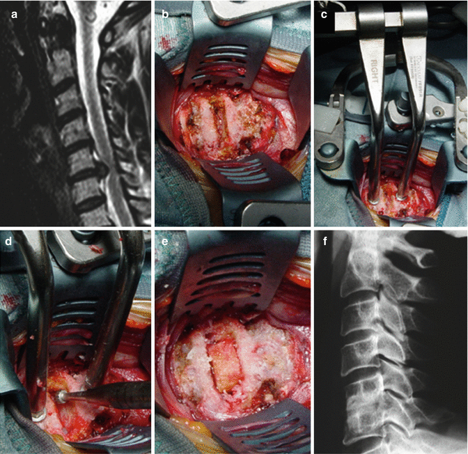
Fig. 9.2
Interbody fusion with Smith-Robinson technique. (a) Preoperative midsagittal T2-weighted MRI scan discloses C5-C6 disc herniation with severe compression of the spinal cord. (b) After exposure of the cervical spine the anulus fibrosus is incised and the anterior longitudinal ligament is removed at the C5-C6 level. (c, d) The disc space is opened with Caspar distraction pin device and the discectomy is completed microsurgically. (e) After complete removal of soft disc herniation, a tricortical autologous iliac crest graft is tapped into place and the distraction is released. (f) Lateral x-ray obtained 6 months after surgery demonstrated solid fusion between C5 and C6
Cloward Technique
This technique increases the space available while the surgeon perform ostheophytes removal and consists of drilling a circular opening in the region of the intervertebral disc, into which a dowel of bone is inserted (Fig. 9.3). After a standard discectomy, measure is taken and the drill depth is determined. The hand-held drill with a guard is applied to the midpoint of the cervical motion segment and drilling of the adjacent vertebral bodies to the desired depth is performed. The guard prevents penetration of the posterior cortical bone, which is removed with high-speed drill and rongeurs. The decompression is completed with microsurgical removal of the PLL, osteophytes and disk herniation, when present. A slightly larger, cylindrical, bicortical autologous dowel graft is harvested from the anterior iliac crest and impacted into the drilled defect. The limitations of this approach consist in the decreased compression strength of the bicortical graft, the extensive exposure of cancellous bone and the impossibility to perform multilevel contiguous fusions [12]. Due to these limitations, the Cloward’s technique is nowadays seldom utilized.
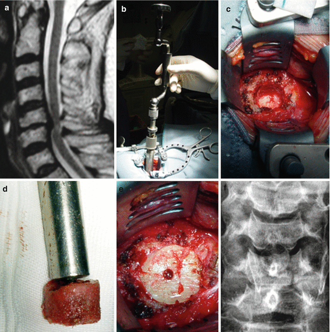
Fig. 9.3
Interbody fusion with Cloward technique. (a) Preoperative midsagittal T2-weighted MRI scan discloses C5-C6 spinal cord compression with high signal changes in the spinal cord as a result of herniated disc associated with osteophytes. (b) After standard discectomy, a hand-held drill is used to drill the adjacent vertebral bodies. (c) The remaining cortical bone is removed microsurgically. (d, e) A bicortical dowel graft of iliac crest bone is tapped into place. (f) Antero-posterior x-ray obtained 6 months after surgery discloses fusion between C5 and C6
Corpectomy
When a corpectomy is planned, the anterior longitudinal ligament over the vertebral body to be resected and over the contiguous portions of the adjacent upper and lower vertebrae is removed (Fig. 9.4). Anterior osteophytes are resected to ensure that the cervical plate will lie flat on the vertebral body. The discetomies above and below the chosen corpectomy site are performed first to evaluate the depth of the spinal canal and to carefully define the limits of bony resection. The majority of the vertebral body is removed under direct visualization to maintain midline orientation and obtain a symmetrical bony decompression. We use a 6-mm cutting burr to rapidly remove the vertebral body to the posterior cortical margin. The location and course of the vertebral arteries is noted on preoperative CT and MRI scans and the distance separating them is measured. Generally, the mean distance separating the medial borders of the transverse foramina is approximately 26–30 mm [22]. According to these findings, we do not extend bony decompression more than 10 mm from the midline to avoid injury of the vertebral artery. Under microscopic view the posterior cortex is removed with a 2-mm Kerrison pouch. The posterior osteophytes are resected with an up-going curette and Kerrison pouch. A matchstick-type burr is used to resect completely the cartilage from the endplates of edjacent vertebrae while maintaining the bony endplate to prevent graft subsidence. The PLL is entered with a nerve hook, incised and widely removed using kerrison pouch. Bone wax is avoided because it prevents bony fusion .
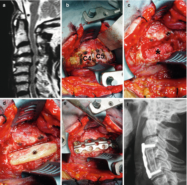
Fig. 9.4
Interbody fusion after corpectomy. (a) Preoperative midsagittal T2-weighted MRI scan shows cervical spine stenosis C3-C5 and kyphotic deformity. (b, c) After wide exposure of midportion of the cervical spine, discectomies are performed above and below the corpectomy site and the vertebral bodies of C4 and C5 are removed using high speed drill. The posterior longitudinal ligament and the osteophytes are resected exposing the decompressed dura (asterisk) (d, e) a tricortical iliac crest graft slightly oversized is tapped into place under distraction and internal fixation is obtained with plate and screws. (f) Lateral x-ray obtained 6 months after surgery demonstrates solid fusion and restoration of focal lordosis
Bony Reconstruction with Autograft
Autologous bone grafts can be classified according to tissue composition (cortical, corticocancellous, cancellous), anatomic origin (iliac crest, rib, fibula) and blood supply (vascularized, nonvascularized). Autografts such as fibula and tricortical iliac crest graft bear the mechanical compression loads applied to the anterior column of the spine due to their strength from their cortical bone composition [27]. These grafts can be fashioned as tricortical or bicortical struts or dowel according to the technique used. Rib grafts are weak mechanically because provide a limited volume of bone and are characterized by a thin cortex. While autologous rib grafts can be used as source of cancellous bone or can wired to the occiput during an occipitocervical fixation, they are not generally used for bony reconstruction of the cervical spine. Vascularized cortico-cancellous autograft such as vascularized fibula grafts are used in irradiated and devascularized fusion beds. The main limitation of fibula graft is the mismatch of the densities with that of the vertebral body with resultant penetration of the fibula through the vertebral body, i.e. the “pistoning effect”. For bony reconstruction of the cervical spine we use autologous bone graft obtained from the anterior iliac crest. Surgical technique plays a pivotal role to ensure proper bone healing and reducing postoperative complications [29]. The bone graft is harvested after the anterior approach and stored in saline-soaked sponges until used. A short (5 cm) skin incision is made parallel to the anterior iliac crest starting at least 2 cm behind and lateral the anterior superior iliac spine (ASIS) (Fig. 9.5). A limited use of electrocautery is required during superiosteal dissection in order to avoid injuring the ilioinguinal, iliohypogastric and lateral femoral cutaneous nerves, which course along the medial surface of the ileum. Care is taken to cut through the fascia avoiding the muscles. The periosteum of the iliac bone is progressively elevated from the inner and outer bone surfaces with a Cobb periosteal elevator. A bone graft of the measured size is harvested from the ilium using a single-bladed oscillating saw under irrigation for tricortical or, more rarely, bicortical bone. The graft site is measured with caliper and the graft is cut slightly oversized (2–3-mm longer than the rostrocaudal length of the corpectomy). Both ends of the graft are flat-surfaced to increase the surface area for fusion. It is our preference to exert distraction of the cervical spine by having the assistant pull on the angle of the mandible on the long axis of the patient’s body. Alternatively, distraction can be obtained by placing distracting pins into the vertebral bodies above and below the corpectomy site. Excessive distraction due to a graft that is to long for the length of the vertebrectomy should be avoided because can lead to postoperative interscapular pain. When the anterior decompression is performed using the Cloward approach, a hand-held drill with a guard is applied on the lateral surface of the ilium at least 2 cm behind the anterior superior iliac spine and a bicortical, cylindrical dowel graft is harvested (Fig. 9.6).