♦ Preoperative
Operative Planning
- Review patient’s history for bone metabolic disease, osteoporosis/osteopenia
- Review patient’s history for diabetes, smoking, and other factors that may affect fusion success rate and therefore intraoperative and postoperative management
- Review preoperative and/or intraoperative imaging to help determine dimensions of area to be instrumented
- Anticipated length of plate to be used
- Anticipated length of screws to be used based on size of vertebral body
- Anticipated length of plate to be used
Equipment
- Select anterior cervical plating system (multiple options of each type)
- Constrained plate systems
- Semiconstrained plate systems
- Dynamic plate systems
- Constrained plate systems
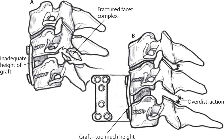
♦ Intraoperative (Fig. 98.1)
Positioning
- Maintain head in neutral position as head turn may lead to unintended fixation in rotated position
Exposure, Decompression, and Reconstruction
- As per primary procedure
- Remove/reduce anterior osteophytes
- Osteophyte rongeur
- Drill (caution to protect surrounding soft tissue structures to limit chances of injury if drill “kicks”)
- Osteophyte rongeur
- Helpful to know width of plate to be inserted
Plate Selection/Placement
- Measure length of area to be spanned by plate (top of superior graft to bottom of inferior graft)
- Use that length to direct plate selection
- Measure distance between bottom of top screw hole to top of bottom screw hole
- Use shortest plate than allows this dimension to allow plate to fully span graft(s)
- Measure distance between bottom of top screw hole to top of bottom screw hole
- Plate may need contouring to match surface of spine (e.g., degree of lordosis)
- If plate contoured, make sure to recheck measurements as length can change
- Carefully adjust retractors to place plate with direct vision and avoidance of soft tissue injury
- Consider use of temporary plate-holding pins
- Verify plate position with x-ray/fluoroscopy
Screw Selection/Placement
- Screw size based on local anatomy, preoperative imaging, and intraoperative imaging
- Most systems do not require bicortical purchase (although may still be
- Rostrocaudal screw angle (Figure 98.1)
- May be directed by plating system
- Should also reflect local anatomy and intraoperative imaging
< div class='tao-gold-member'>
Fig 98.1 Schematic of (A) inadequate graft height with secondary facet fracture, and (B) graft overdistraction.
Only gold members can continue reading. Log In or Register to continue
Stay updated, free articles. Join our Telegram channel
- May be directed by plating system

Full access? Get Clinical Tree


