♦ Preoperative
Imaging
- Magnetic resonance imaging to assess spinal cord compression
- Plain x-rays to evaluate alignment
- Dynamic, flexion/extension radiographs can be helpful in the evaluation of flexibility of the spine and ability to restore alignment without osteotomies
- Computed tomography with sagittal reconstructions to evaluate alignment and to visualize limitations of possible exposure (level of sternal notch, angu-lation of disc spaces, and depth of spine from skin surface)
Preoperative Care
- Somatosensory evoked potentials/motor evoked potentials may be useful
Equipment
- Self retaining anterior cervical retraction system (if possible, obtain two sets or longer blades)
Operating Room Set-up
- Somatosensory and motor evoked potential monitoring (optional)
- Fluoroscopy (consider draping into field)
- Balanced microscope
Positioning
- Supine on operating table
- Head fixed in Mayfield head holder if destabilizing osteotomies/anterior releases are planned
♦Intraoperative
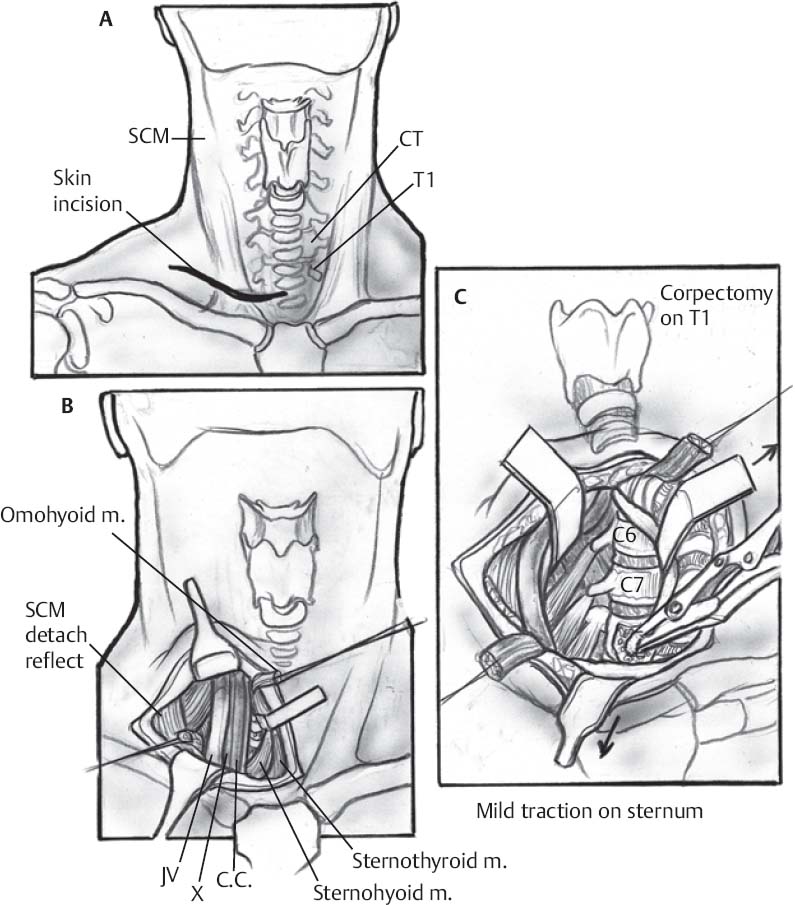
Exposure (Fig. 97.1)
- Horizontal incision in lowest skin crease (at least 3 cm above sternal notch) on the right
- Divide platysma from midline to medial border of sternocleidomastoid.

Fig 97.1 Anterior cervicothoracic junction approach. C.C., common carotid artery; JV, jugular vein; SCM, sternocleidomastoid muscle.
Only gold members can continue reading. Log In or Register to continue
Stay updated, free articles. Join our Telegram channel

Full access? Get Clinical Tree


