(1)
Marina Spine Center, Marina del Rey, CA, USA
1.
Position the patient supine on the table, the lower lumbar spine at the level of the kidney rests.
2.
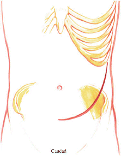
Make a lower abdominal pararectus incision through the skin and subcutaneous tissue (Fig. 28.1). The most immediate layers are the external oblique with its transition into the linea semilunaris leading to the fascia of the rectus sheath. The linea semilunaris is composed of the aponeurosis of the three layers of the abdominal musculature and their fascia.


Fig. 28.1
Position the patient supine on the table with the lumbar spine over the kidney rests. Make a lower abdominal pararectus incision through the skin and subcutaneous tissue. The most immediate layers are the external oblique with its transition into the linea semilunaris leading to the fascia of the rectus sheath. The linea semilunaris is composed of the aponeurosis of the three muscle layers of the abdominal musculature and their fascia
3.
Incise the fibers of the external oblique, the internal oblique, and the small thin layer of transversus abdominis muscles lateral to the semilunaris in line with the skin incision.
4.
Identify the transversalis fascia. The transversalis fascia is the internal investing fascial layer of the abdominal cavity. Dissect on the outer surface of this transversalis fascia to the edge of the rectus sheath. At this point the transversalis fascia splits to form the lamina of the rectus sheath. The posterior lamina of the rectus sheath forms the endoabdominal fascia in this area.
5.
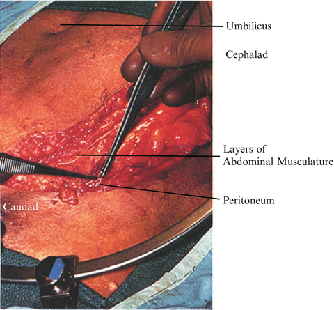
Incise the transversalis fascia lateral to the linea semilunaris carefully and identify the peritoneum through the incision (Fig. 28.2).


Fig. 28.2
Incise the external oblique, internal oblique, and transversus abdominis muscles, lateral to the semilunaris in line with the skin incision. Identify the transversalis fascia. Incise the transversalis fascia lateral to the semilunaris and identify the peritoneum through the incision. Dissect the peritoneum with a sponge or gloved hand off the undersurface of the transversalis fascia and open the abdominal wall after the peritoneum has been identified and cleared from the abdominal wall. The left rectus muscle may be divided for increased ease of retraction. Note: The peritoneum may be identified and dissection begun under the rectus sheath, but it is easier to find it laterally
6.
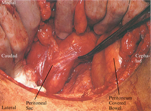
Begin the incision in line with the skin incision, dissecting the peritoneum with a sponge or gloved hand off the undersurface of the transversalis fascia. Open the abdominal wall after the peritoneum has been identified and cleared (Fig. 28.3).


