(1)
Marina Spine Center, Marina del Rey, CA, USA
The anterior lateral approach is medial to the sternocleidomastoid muscle and lateral to the carotid sheath. For approaches to C1, C2, and C3, in which the head should not be turned or rotated, and for surgeons who do not feel comfortable with the neurovascular structures medial to the carotid sheath, this approach offers a relatively bloodless field.
1.
Position the patient supine. Use the neurosurgical head-holding frame with skeletal skull fixation for better exposure and stability.
2.
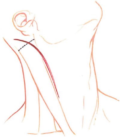
Incise the skin and subcutaneous tissue beginning on the medial border of the sternocleidomastoid muscle at the superior edge of thyroid cartilage and extend this cephalad to the mastoid process and curve posteriorly across the process [1] (Fig. 6.1).


Fig. 6.1
With the ear lobe retracted medially by a stay suture, make a longitudinal incision along the anterior border of the sternocleidomastoid muscle, posteriorly curving across the mastoid process. The dotted line is the incision in the insertion of the sternocleidomastoid muscle
3.
Open the external investing fascia along the line of the incision. Place superflcial spring retractors and obtain hemostasis. Avoid the cutaneous sensory nerves crossing the sternocleidomastoid muscle.
4.
Get Clinical Tree app for offline access

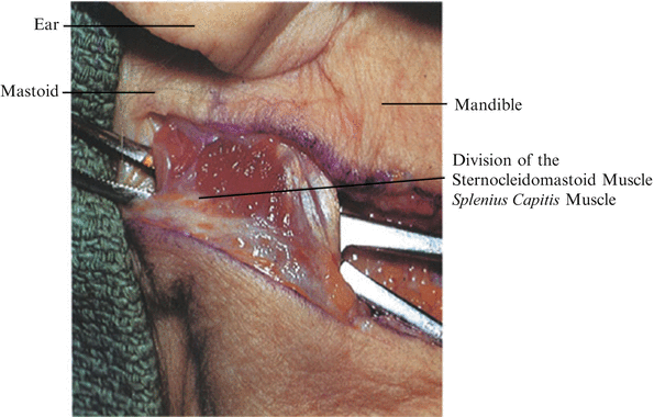
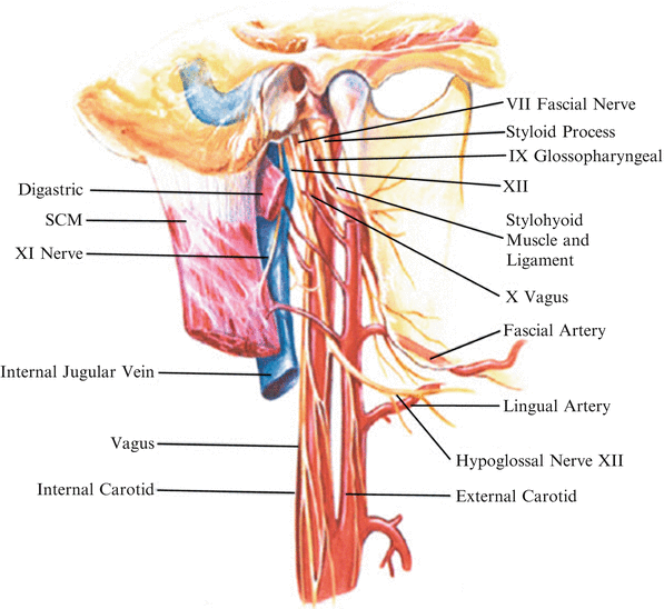
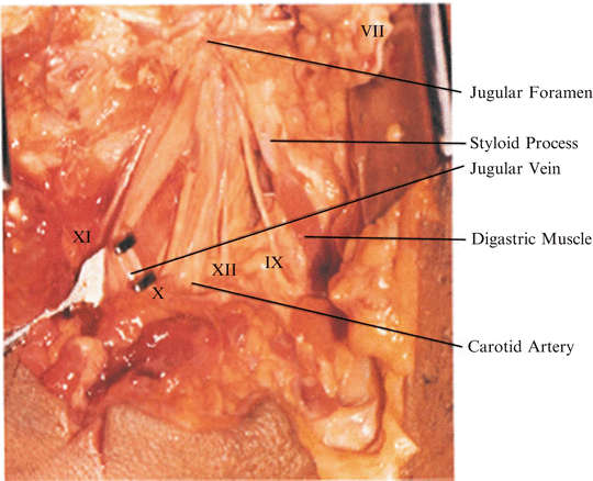
Palpate the transverse process of Cl and the mastoid process. Delineate the medial edge of the sternocleidomastoid muscle with scissors (Fig. 6.2). Incise the fascia and sternocleidomastoid muscle at the insertion of the sternocleidomastoid and mastoid portion of the splenius capitus on the mastoid process. Leave a fascial edge suitable for reattachment. The splenius capitis is deep in the wound. Incise the superficial medial portion. Remember that the mastoid process is just posterior to the styloid process (Fig. 6.3). Between the two is the stylomastoid foramen from which exits the facial nerve (Fig. 6.4). The jugular bulb enters the skull anteromedial to the mastoid and stylohyoid processes. The jugular foramen from which exit cranial nerves XI (accessory), X (vagus nerve), IX (glossopharyngeal nerve), and XII (hypoglossal) is medial to the jugular bulb (Fig. 6.3). The accessory nerve courses laterally under the internal jugular vein to reach the sternocleidomastoid muscle (Fig. 6.5).


Fig. 6.2
After palpation of the transverse process of C1 and the mastoid process, delineate the medial and lateral edges of the sternocleidomastoid with scissors. Incise the fascia and the sternocleidomastoid muscle and the mastoid portion of the splenius capitis muscle on the mastoid process. Leave a fascial edge suitable for reattachment

Fig. 6.3
The structures exiting the jugular bulb are the hypoglossal nerve, vagus nerve, spinal accessory nerve, and glossopharyngeal nerve. The jugular bulb is the entry point of the jugular vein and carotid artery. Cranial nerve VII exits from the stylomastoid foramen. Sternocleidomastoid muscle (SCM)

Fig. 6.4


After identification of the mastoid process, remember that just medial to the mastoid process is the styloid process. Between the two is the stylomastoid foramen, from which exits cranial nerve VII. Un-needed cephalad dissection or too vigorous retraction may damage cranial nerve VII at this point. The jugular bulb enters the skull anteromedial to the mastoid and stylohyoid processes. Medial to the jugular bulb is the jugular foramen from which exit cranial nerves XI (accessory), X (vagus nerve), XII (hypoglossal nerve), and IX (glossopharyngeal nerve). The accessory nerve courses laterally over the internal jugular vein to reach the sternocleidomastoid muscle. The other cranial nerves course medially in the medial musculovisceral column
Stay updated, free articles. Join our Telegram channel

Full access? Get Clinical Tree


