(1)
Marina Spine Center, Marina del Rey, CA, USA
1.
The standard approach is from the left side to avoid the recurrent laryngeal nerve. Rotate the head to the right and inflate the cervical pillow to support the head and neck. The necessity for intraoperative traction is determined by the pathologic lesion. When necessary, use Gardner Wells tong traction rather than the halter.
2.
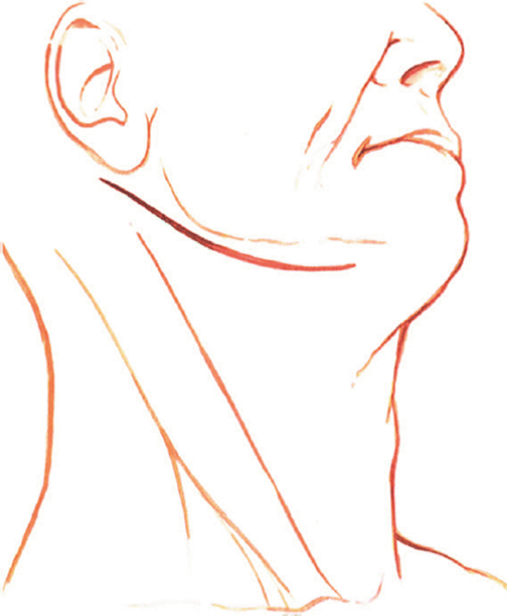
Make the skin incision transversely starting one fingerbreadth from the midline below the mandible and proceed laterally, curving around the angle of the mandible posteriorly across the mastoid process (Fig. 5.1). For a pathologic process that extends to C3 or below, make the skin incision along the anterior border of the sternocleidomastoid muscle, extending it cephalad and curving in a similar manner across the mastoid process.


Fig. 5.1
The skin incision is made under the angle of the mandible from posterior sternocleidomastoid muscle toward the midline
3.
Incise the layer of the platysma muscle, inserting small spring retractors in the subcutaneous tissue. Open the platysma muscle along the line of the incision and retract it in a similar manner. The greater auricular nerve and anterior cervical nerve are branches of the ansa cervicalis that course around from the posterior edge of the sternocleidomastoid muscle, crossing the muscle for distribution to the auricular and mandibular skin. Preserve these nerves when possible [1].
4.
Identify the medial border of the sternocleidomastoid muscle. To facilitate this identification use scissors to incise the superficial investing fascia. To locate the carotid sheath, palpate the carotid pulse. Use finger dissection and blunt right-angle blade retraction to develop the incision medial to the sternocleidomastoid muscle and the carotid sheath and lateral to the musculovisceral column (strap muscles, esophagus, and trachea).
5.
If needed for additional exposure, divide the anterior third of the insertion of the sternocleidomastoid muscle from the mastoid process. Leave sufficient fascial tissue to reattach the muscle when closing the wound [2].
6.
Palpate the carotid pulse and deepen the incision medial to the carotid sheath and the sternocleidomastoid muscle through the middle cervical fascia with blunt, longitudinal finger dissection and scissors.
7.
The neurovascular structures crossing from lateral to medial are found in this layer.
8.
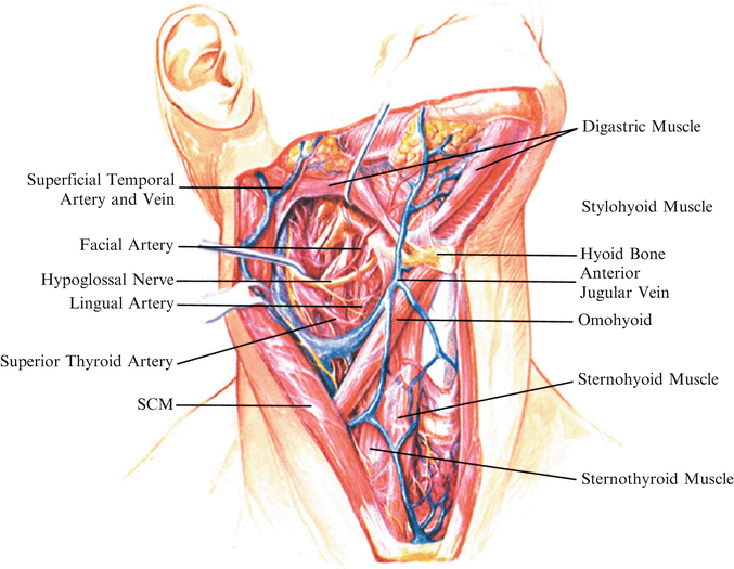
Identify the superior thyroid artery and vein. The superior thyroid artery arises from the external carotid artery at approximately the level of the hyoid bone. It crosses through the carotid triangle, arches deep to the strap muscles, and enters the lateral superior aspect of the thyroid gland. Retract the superior thyroid artery and vein inferiorly (Fig. 5.2).


Fig. 5.2
Dissection of the cephalad portion of the anterior neck shows the carotid triangle area, which is bordered superiorly by the posterior belly of the digastric muscle, inferiorly by the omohyoid muscle, and posteriorly by the sternocleidomastoid muscle. The digastric muscle lies inferior to the mandible and extends from the mastoid process to the symphysis mente. The sling around the tendonous midportion of the digastric muscle separates its posterior from its anterior belly, and holds the midportion of the muscle of the comu of the hyoid bone. The stylohyoid muscle (retracted by hook) lies anterior and superior to the posterior belly of the digastric, passing from the styloid process to the hyoid bone. Within the carotid triangle the lingual artery, the facial artery, and the hypoglossal nerve course from lateral to medial. The hypoglossal nerve is usually retracted cephalad, for the cephalad approach to C1–C2, and the lingual and facial arteries are sacrificed. Caution: Always identify the hypoglossal nerve before any ligation of structures in this area
9.
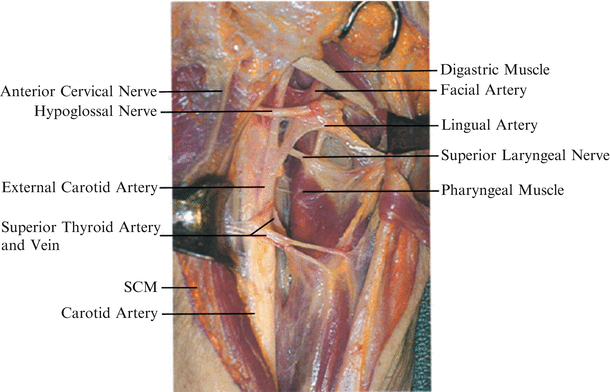
Identify and retract the hypoglossal nerve (Fig. 5.3). The hypoglossal nerve is found passing from lateral to medial superficial to the external carotid, lingual, and facial arteries. The hypoglossal nerve exits the skull in close proximity to the vagus nerve and courses between the internal carotid artery and internal jugular vein, becoming superficial at the angle of the mandible. After its usual point of identification over the arteries, it passes deep to the tendon of the digastric muscle and stylohyoid muscle for distribution to the muscles of the tongue. The hypoglossal nerve is retracted cephalad, and the superior thyroid artery and vein are usually retracted caudad.


