(1)
Marina Spine Center, Marina del Rey, CA, USA
1.
Position the patient supine on the operating table. Turn the head to the right.
2.
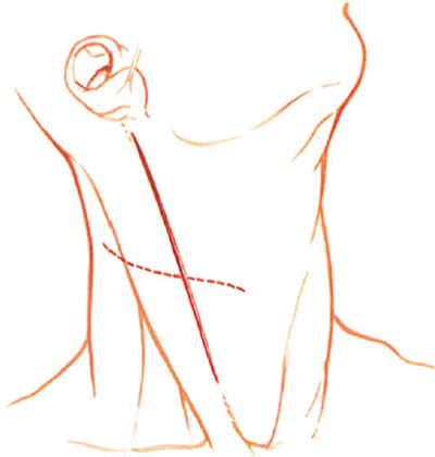
Usually, the incision goes from the midline to the medial sternocleidomastoid muscle (SCM). After the transverse skin incision (Fig. 7.1), at the appropriate level, dissect through subcutaneous tissue to the platysma muscle. Open the platysma muscle in the line of its fibers when possible. Elevate the platysma muscle with Adson pick-ups and open carefully. Beware of damage to veins and the sternocleidomastoid muscle [1 , 2]. Insert spring retractors.


Fig. 7.1
The more cosmetically suitable transverse incision is made at the appropriate level and should allow exposure of as many as two discs and three vertebrae. A vertical incision can be used for an even greater exposure
3.
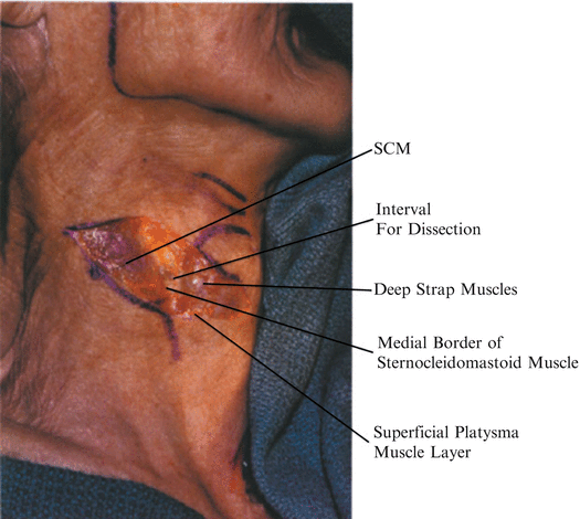
Open the superficial, cervical fascia and identify the medial border of the sternocleidomastoid muscle [3]. “The first key to successful exposure is adequate identification of the medial border of the sternocleidomastoid so that may be retracted laterally [4]. With identification of this medial border, bluntly develop the interval between the sternocleidomastoid muscle and the sternohyoid and sternothyroid muscles (Figs. 7.2, 7.3). Retract the posterior cutaneous nerves. Bluntly dissect and spread the soft tissue vertically in this interval.


Fig. 7.2
After the skin incision, dissect through subcutaneous tissue to platysma muscle, which in this specimen is very thin. Open the platysma muscle carefully in line with its fibers. Beware of damage to veins and the sternocleidomastoid muscle (SCM). Insert spring retractors

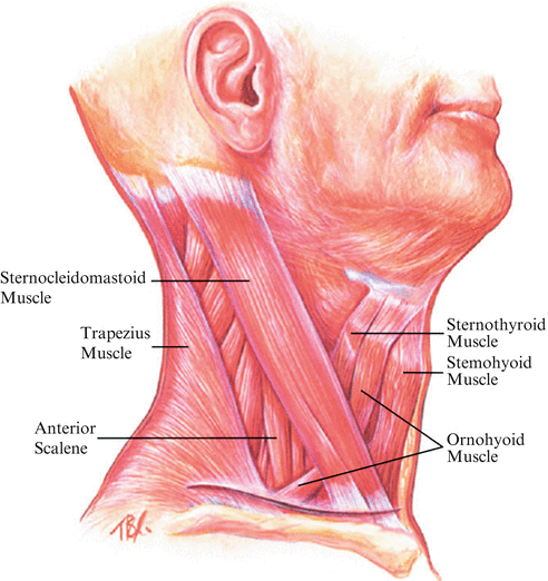
Fig. 7.3
The key to the dissection at this point is to identify the medial border of the sternocleidomastoid muscle. With lateral retraction of the sternocleidomastoid, the interval between this muscle and the medial strap muscles is delineated
4.
Get Clinical Tree app for offline access

Retract the sternocleidomastoid laterally and the strap musculature medially with angled retractors. Identify the middle cervical fascia. The omohyoid muscle crosses from proximal medial to lateral distal through the middle cervical fascia at C6–C7 (Figs. 7.3, 7.4, 7.5). (The omohyoid arises from the scapula. Its inferior belly crosses ventromedially through a sling of deep cervical fascia that attaches to the clavicle. The muscle passes under the sternocleidomastoid muscle close to the lateral border of the sternohyoid muscle and inserts into the body of the hyoid bone.) Retract the omohyoid; when necessary divide it laterally.


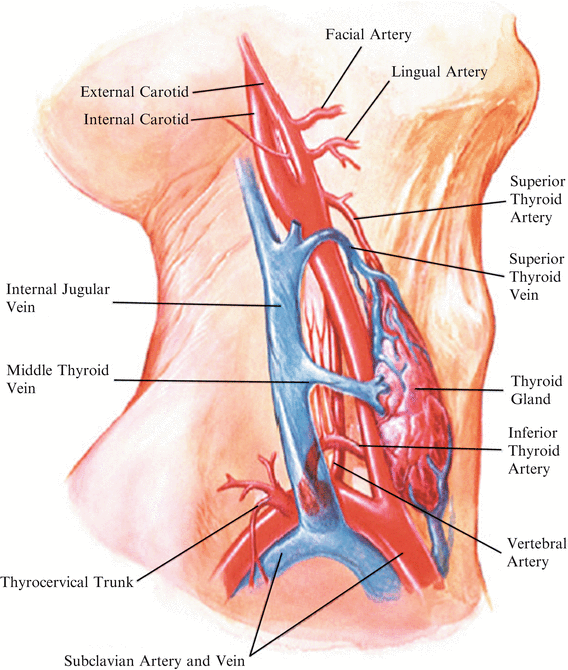
Fig. 7.4


After retracting the sternocleidomastoid muscle laterally and the strap musculature medially, the arteriovenous structures of the middle cervical fascial layer must be identified. Palpate the carotid pulse. Open the midline cervical fascia medial to the carotid artery. Ligate and tie the medial thyroid vein. Retract the superior thyroid artery cephalad and the inferior thyroid artery caudad to expose the midcervical spine
Stay updated, free articles. Join our Telegram channel

Full access? Get Clinical Tree


