Antiplatelet- and Anticoagulation-Associated Intracranial Hemorrhage

4 Antiplatelet- and Anticoagulation-Associated Intracranial Hemorrhage


Fred Rincon, Andres Fernandez, and Stephan A. Mayer


Antiplatelet- and anticoagulation-associated intracranial hemorrhages are a neurologic emergency. As the population ages, anticoagulation-associated intracranial hemorrhage is expected to increase in frequency.


Case Example


A 39-year-old right-handed Hispanic man with a history of recent mechanical aortic valve replacement (AVR) was admitted to the hospital for an evaluation of his chest wound infection. The day after admission his mental status worsened, and the physical examination revealed a new right hemiparesis with left-gaze deviation.


Questions


  • What was the time of onset? (When was the patient last seen normal?)
  • Is the patient on antiplatelet or anticoagulant medications?
  • If the patient is having an ischemic stroke, should we administer intravenous (IV) tissue plasminogen activator (tPA) or consider any other intervention?
  • In case of coagulopathic ICH, when can we restart anticoagulation in this patient with a mechanical aortic valve?

Urgent Orders


  • Assess for intubation (airway, breathing, circulation [ABCs]).
  • Perform head computed tomography (CT) without contrast.
  • Check coagulation and platelet profiles.
  • Order appropriate reversal agents as soon as possible if the patient has received anticoagulant or antiplatelet medication, or has abnormal coagulation or platelet studies.

History and Examination


History


  • Assess antiplatelet use (aspirin, clopidogrel, dipyridamole, ketorolac, ticlopidine, GPIIb/IIIa medications), anticoagulation medications (low molecular weight heparin, unfractionated heparin, direct thrombin inhibitors, or warfarin), as well as when each medication was last administered.
  • Assess for previous stroke, baseline functional status, history of hypertension (HTN), dementia, liver disease, or renal failure.
  • Check for history of tobacco, cocaine, amphetamine, and over-the-counter drug use.
  • Assess for dysfunctional platelets (renal disease) or intrinsic coagulopathy (hemophilia, von Willebrand disease).

Physical Examination


Note blood pressure (BP); fever should be aggressively treated.


Neurologic Examination


Differential Diagnosis


  1. Intracranial hemorrhage (intracerebral hemorrhage [ICH], subarachnoid hemorrhage [SAH], subdural hematoma [SDH], or epidural hematoma [EDH]). Although SAH is often accompanied by “the worst headache in my life” and SDH/EDH by history of head trauma, they may occur spontaneously, especially in the setting of coagulopathy. A growing source of ICH is vitamin K inhibitor use, which accounts for ~10 to 15% of ICH.1 Coagulopathic bleeds are often multifocal. Common sites include the cerebellar vermis and hemorrhage into previously ischemic tissue. Patients with warfarin- or antiplatelet-associated ICH have increased mortality and increased risk of ICH expansion compared with noncoagulopathic ICH.

    • Long-term anticoagulation may increase the risk of ICH by 10-fold, and the annual rate of ICH for patients on warfarin is ~1%.2 The main risk factors for warfarin-associated ICH include age, hypertension, intensity of anticoagulation, concomitant aspirin use, cerebral amyloid angiopathy, and leukoaraiosis.3 The risk of ICH increases with international normalized ratio (INR) values over 3.5 to 4.5 and nearly doubles for each increase of 0.5 point over 4.5.1 Despite this increased risk with increasing INR, most anticoagulation-associated bleeds occur with an INR in the recommended therapeutic range.
    • Antiplatelet agent use has a risk of symptomatic bleeding complications, but there is meager evidence implicating the use of a single antiplatelet agent as a risk factor for ICH.4,5 However, the combination of antiplatelet agents such as aspirin (ASA) and ADP-receptor blockers such as clopidogrel carries a higher incidence of ICH. Other antiplatelet agents such as the glycoprotein receptor blocking agents (GPIIb/IIIa) produce hematologic abnormalities that may put the patient at risk of bleeding as well.

  2. Ischemic stroke. In patients with a history of a cardiac disease (recent open heart surgery and mechanic AVR) or atrial fibrillation (AF), ischemic stroke is a plausible diagnosis. The etiology of ischemic stroke in these settings is usually embolic (thrombus or septic emboli). Hemorrhage into an area of ischemic infarction occurs when vessel walls are damaged by ischemia, and blood then extravasates into the brain parenchyma. The transformation requires sufficient time for an ischemic lesion to develop and then partial or total reperfusion with restoration of blood flow through the vessel or by collateralization. Large infarct size, older age, hyperglycemia, sustained hypertension, thromboembolic mechanism (as opposed to penetrator occlusion), and preexisting micro-hemorrhages on magnetic resonance imaging (MRI) have been identified as risk factors for hemorrhagic conversion of an infarct. Small asymptomatic petechiae are less important than frank hematomas, which may be associated with neurologic decline. In general, spontaneous hemorrhagic conversion after ischemic stroke occurs in 0.6 to 5% of patients admitted to the hospital. Management of spontaneous hemorrhagic transformations depends on the amount of blood and clinical symptoms. According to the European Cooperative Acute Stroke Study (ECASS) criteria, hemorrhagic conversion of an infarct is graded as seen in Table 4.1.6 Patients exposed to recombinant tissue plasminogen activating factor (rtPA) for ischemic stroke or myocardial infarction have a risk of symptomatic ICH of 6 to 7% and 0.2 to 1.4%, respectively. ICH following fibrinolysis has a 30-day mortality rate of 60%.2


    Table 4.1 Hemorrhagic Conversion of an Infarct
    HI 1 Small petechial hemorrhage
    HI 2 Confluent petechial hemorrhage
    PH 1 Hematoma in <30% of the infracted area with minimal mass effect
    PH 2 Hematoma in >30% of the infracted area with significant space occupying effect

    Abbreviations: Hl, hemorrhagic infarct; PH, parenchymal hematoma.


    Data from: Molina CA, Alvarez-Sabin J, Montaner J, et al. Thrombolysis-related hemorrhagic infarction: a marker of early reperfusion, reduced infarct size, and improved outcome in patients with proximal middle cerebral artery occlusion. Stroke 2002;33(6):1551–1556.


    • Seizures. New onset seizures with focal neurologic deficits (Todd’s paralysis) may be considered as part of the differential diagnosis. It is important to mention that seizures may accompany some stroke types, especially hemorrhagic ones.

Life-Threatening Diagnoses Not to Miss


  • Coagulopathy associated hemorrhage as urgent reversal can improve outcome.
  • Ischemic stroke that may qualify for thrombolysis (see Chapter 6).

Diagnostic Evaluation


  • Laboratory studies. Review prothrombin time/partial thromboplastin time/international normalized ratio (PT/PTT/INR), complete blood count (CBC), type and hold, troponin, chemistry panel, magnesium, calcium, phosphorus, and liver function test for all patients. In selected patients check D-dimer, fibrinogen, toxicology screen, and bleeding time.
  • Imaging studies. Order head CT without contrast, consider CT angiogram, MRI, MR angiogram (see Chapter 3).
  • ICP monitoring may be helpful in patients with decreased level of consciousness or IVH.
  • Continuous blood pressure and cardiac rhythm monitoring should be performed at least for the initial 24 hours because of the high rate of cardiovascular instability.

Treatment


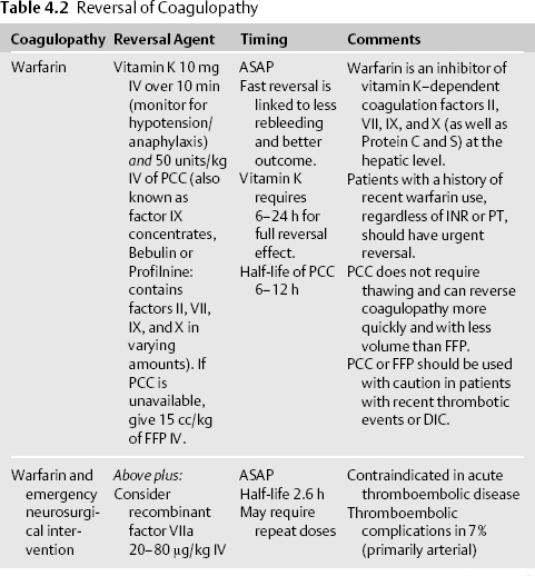
Reversal Suggestions7 (Table 4.2)


Stay updated, free articles. Join our Telegram channel

Aug 30, 2016 | Posted by in NEUROSURGERY | Comments Off on Antiplatelet- and Anticoagulation-Associated Intracranial Hemorrhage

Full access? Get Clinical Tree

Get Clinical Tree app for offline access