(1)
Marina Spine Center, Marina del Rey, CA, USA
1.
Position the patient’s head in the self-retaining neurosurgical head fixation device that is attached to the table. Attach the drapes to the patient’s neck with stay sutures. Neck flexion will increase exposure, but flexion is limited by the type of pathology present, usually to a neutral, slightly flexed position. With instability problems, confirm the position of the spine with X-ray.
2.
Incise the midline skin and subcutaneous to fascia and obtain hemostasis with rapid application of hemostats and electrocautory. Insert self-retaining retractors.
3.
Deepen the incision with the cautery knife, staying in the thin white median raphe, and avoid cutting muscle tissue. The median raphe of the cervical spine is a tortuous structure; it does not follow a straight midline incision. Open the median raphe to the spinous processes of C2 and C3 and the occiput. In children, to avoid spontaneous fusion at adjacent levels, including the occiput, expose no spinal levels unnecessarily.
4.
Expose the spinous processes with a #15 blade or cutting cautery on their bulbous bifid tips. The ligamentous attachments to C2 are very prominent. Identify any spina bifida of the cervical spine on the preoperative X-ray and at surgery. Insert the Cobb elevator, first facing up to elevate the tip subperiosteally, then facing down to complete the subperiosteal elevation medial to lateral approximately 1 inch at each level. At levels below C2, identify the medial edge of the facet joint at the base of the lamina and pack at each level as it is exposed. When necessary, expose the occiput with elevators. Insert self-retaining retractors to expose the base of the skull and the dorsal spine of C2. The area in between will contain the ring of C1. Many times this is very deep compared with C2.
5.
Maintain firm lateral traction on the wound. With a sharp Cobb elevator, feel for and identify the posterior tubercle of C1 longitudinally in the midline. Begin the subperiosteal dissection to bone.
Caution: Often C1 is very thin, and direct pressure can fracture it or cause the surgeon to slip off the ring and penetrate the atlanto-occipital membrane. Elevation on this ring can be very dangerous if there is subluxation with constriction of the posterior dura under this ring. The dura may be vulnerable on both the superior and inferior edges of the ring of C1.
6.
Get Clinical Tree app for offline access

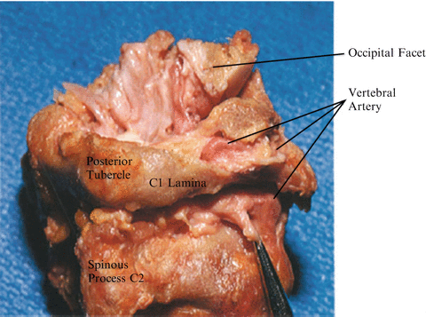
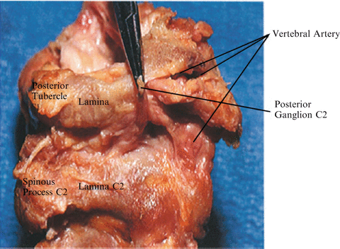
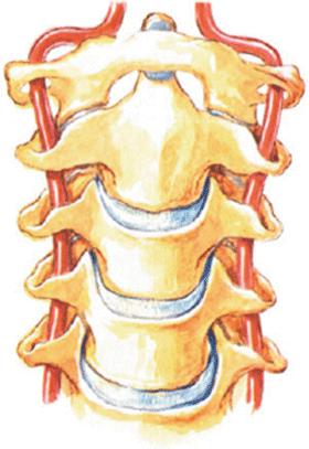
Dissect laterally only approximately 1.5 cm. An important landmark on the ring of C1 laterally is the second cervical ganglion, which lies at approximately 1.5 cm on the lamina of C1 in the area of the groove for the vertebral artery (Figs. 38.1, 38.2). Carefully identify the most medial aspect of the groove for the vertebral artery and vein on the superior border of the C1 ring [1]. The bluish color of the vein is visualized first (Fig. 38.3). By seeing the initial ridge or the vein one can avoid damaging the artery.


Fig. 38.1
A dissected specimen of the C1–C2 articulation emphasizes the two potential areas of damage to the vertebral artery in this area. One is between the C1–C2 articulation in an exposed, posterior lateral position; the other is on the superior ring of C1. This specimen has a bone encasing the vertebral artery. A portion of much of the roof of the tunnel has been removed, emphasizing that when dissecting on the ring of C1, identification of the edge of the groove immediately preceding the vertebral artery allows one to locate the artery before any damage. The specimen does not show the bluish hue of the vessels usually encountered operatively. In the forceps is the posterior ganglion and ramus of C2, which is the warning landmark one reaches laterally before the vertebral artery between C1 and C2

Fig. 38.2
The usual location of the second cervical ganglion medial to the vertebral artery

Fig. 38.3


The vertebral artery enters the foramen transversarium of C6, and courses through each foramen transversarium anterior to the spinal nerve. The foramen transversarium of C1 is in a relatively more posterior position than that of the lower cervical vertebrae. After passing through the foramen transversarium of C1, the vertebral artery courses posteromedially to enter the spinal canal and the foramen magnum
Stay updated, free articles. Join our Telegram channel

Full access? Get Clinical Tree


