Approach to the Sensory Examination
PURPOSE
The main purpose of the sensory examination is to localize neurologic pathology by looking for characteristic distributions of sensory loss.
WHEN TO PERFORM THE SENSORY EXAMINATION
A detailed sensory examination, including testing for pin and vibratory sensation, should be performed on any patient who presents with sensory symptoms, such as numbness or tingling in a part of the body or extremities. A detailed sensory examination should also be performed on any patient with signs or symptoms of any focal disorder of the central of peripheral nervous system, because the finding of associated sensory loss may aid in localization.
In patients without sensory symptoms or other significant focal neurologic symptoms or signs, however, the sensory examination plays a limited role. In these patients, an assessment of distal vibratory sensation usually suffices to confirm normal sensory function.
NEUROANATOMY OF SENSATION
The sensory pathways for the body and the extremities begin in peripheral receptors, travel up the peripheral nerves to the dorsal nerve roots, and then enter the spinal cord. In the spinal cord, the sensory pathways ascend as the spinothalamic tract (mainly subserving pain and temperature sensation; see Chapter 29, Examination of Pinprick Sensation) and the posterior columns (mainly subserving vibration and proprioceptive sensation; see Chapter 30, Examination of Vibration and Position Sensation). After ascending in the spinal cord, these tracts synapse in the contralateral thalamus, where the sensory information is then relayed to the cerebral cortex.
Dermatomes are the areas of skin where the sensation is supplied by a single nerve root. Dermatome charts (Fig. 28-1) summarize the approximate cutaneous sensory territory of nerve root innervation of the body and the extremities. These charts are helpful in localizing patients’ sensory symptoms to the appropriate region when those symptoms are due to a lesion of the nerve root or the spinal cord. The cutaneous distribution of the peripheral nerves themselves (Fig. 28-2) can also be shown graphically and can aid in localization of patients’ sensory symptoms to a particular peripheral nerve lesion.
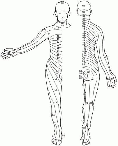 Figure 28-1 Dermatome chart of the anterior and posterior body and extremities. (From Dudek RW. High-yield gross anatomy. Philadelphia: Williams & Wilkins, 1997.) |
EQUIPMENT NEEDED TO TEST SENSATION
256-Hz tuning fork
Safety pin (see Chapter 29, Examination of Pinprick Sensation, for details on the use of a safety pin or appropriate alternative to test pin sensation)Related posts:

Stay updated, free articles. Join our Telegram channel

Full access? Get Clinical Tree


