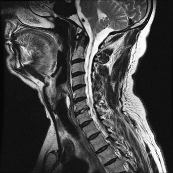
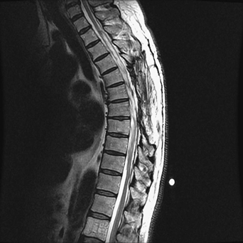
83 A 60-year-old man who had a previous thoracic laminectomy for resection of an arachnoid cyst continued to have progressive myelopathy. T2-weighted sagittal magnetic resonance imaging (MRI) of the spine shows a septated arachnoid cyst spanning from the cervicomedullary junction into the thoracic spine (Figs. 83-1 and 83-2). The cyst is cerebrospinal fluid (CSF) density on MRI. The individual compartments communicate as seen on myelogram (Figs. 83-3 and 83-4). FIGURE 83-1 MRI of the cervical spine demonstrates the arachnoid cyst. Recurrent, enlarging, spinal arachnoid cyst FIGURE 83-2 MRI of the thoracic spine demonstrates the arachnoid cyst.
Arachnoid Cyst
Presentation
Radiologic Findings
Diagnosis
< div class='tao-gold-member'>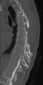
Only gold members can continue reading. Log In or Register to continue


