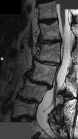
85 An 87-year-old woman presented with chronic low back pain radiating to the posterior thighs. She had two lumbar decompressions and one non-instrumented fusion in the past. In addition, she has had several oil-based myelograms throughout her life. She is neurologically normal. FIGURE 85-1 Sagittal MRI of the lumbar spine shows clumping of the cauda equina. Sagittal and axial T2-weighted magnetic resonance imaging (MRI) of the lumbar spine highlights spondylotic as well as postoperative changes, with notable nerve root clumping (Figs. 85-1 and 85-2). Arachnoiditis
Arachnoiditis
Presentation
Radiologic Findings
Diagnosis
< div class='tao-gold-member'>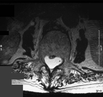
Only gold members can continue reading. Log In or Register to continue


