Wartenberg’s Sign
Active flexion of the terminal phalanges of the four fingers of a paretic hand about a firm object, or against resistance offered by the examiner’s fingers similarly flexed, is followed by adduction, flexion, and opposition of the thumb (Figure 42.1). In a normal extremity, the thumb remains in abduction and extension. A variation is for patient and examiner to hook and pull with only the index fingers; the response is the same.

FIGURE 42.1 AM of thumb (Wartenberg’s sign). A. The patient bends his last four fingers against resistance of four hooked fingers of the examiner. Thumb moves toward palm. Mild spastic paralysis of hand. B. With his fingers hooked over a horizontally fastened rod, the patient is asked to pull it down. Right thumb performs an AM toward the palm. Right-sided spastic hemiplegia. (From Wartenberg R. Diagnostic Tests in Neurology. Chicago, IL: Year Book Medical Publishers, 1953.)
The Trunk-Thigh Sign of Babinski, or Combined Flexion of the Trunk and Thigh
The patient, lying supine with legs abducted, attempts to sit up while holding the arms crossed on the chest. Normally, the legs remain motionless and the heels down. In corticospinal hemiparesis, the hip flexes as the trunk flexes, and there is an involuntary elevation of the paretic limb off the bed (Figure 42.2). The toes may spread out in a fan-like fashion. The normal limb remains on the bed or rises slightly, but not as high as the paretic one. In paraparesis, both legs rise equally. In nonorganic weakness, the normal leg rises and the paretic one does not, or neither leg rises. If the patient tries to sit up with the legs hanging over the edge of the bed, the hip flexes and the knee extends on the involved side. The same phenomenon occurs if the standing patient bends over (Figure 42.3).
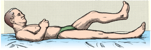
FIGURE 42.2 Trunk-thigh sign in patient with left hemiparesis.
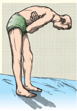
FIGURE 42.3 Combined flexion of the thigh and leg in a patient with left hemiparesis.
The Tibialis Sign of Strümpell
Normally, vigorous flexion of the hip and knee are accompanied by plantarflexion of the foot. In lower-extremity weakness due to a corticospinal tract lesion, voluntary flexion of the hip and knee is accompanied by involuntary dorsiflexion and inversion ofthe paretic foot; there may also be dorsiflexion of the great toe or of all the toes. The patient is unable to flex the hip and knee without dorsiflexing the foot (Figure 42.4). The response is accentuated if the movement is carried out against resistance. The sign may also be elicited by flexing the knee with the patient lying prone (Figure 42.5).
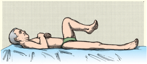
FIGURE 42.4 Tibialis sign in a patient with left hemiparesis.


