Attachment
PARENTAL BEHAVIOR
The goal of reproduction is successful offspring. Parents want offspring who can survive the rigors of the world and produce their own descendents. Many animals produce offspring that require sustained assistance to successfully reach maturity. The particular actions that parents undertake to ensure the growth and survival of their offspring constitute parental behavior.
The extent of parental behavior in the animal kingdom occurs along a spectrum ranging from none to helicopter parents. Female salmons lay hundreds of eggs to be fertilized and then swim away. Humans are at the other end of the spectrum—investing many years and enormous resources, and perhaps hoping to be surrounded by their children until the very end.
Females in the animal kingdom do most of the parenting, although there are some exceptions. It is generally believed that males seek to fertilize as many eggs as possible, whereas females seek to successfully raise the few they sire. Parental behavior constitutes any behavior that the parent does for the offspring. For example, a pregnant dog will build a nest a day or two before giving birth. After the delivery, she will lick them clean, eat the placentas, feed them, and keep them warm (and for all this she is called a bitch). Additionally, she will aggressively defend the pups against any suspicious intruders.
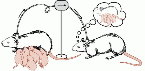
The onset of maternal behavior is remarkably precise. An inexperienced mother must immediately perform a full range of new behaviors without much room for error. How does this happen? Terkel and Rosenblatt established that there must be something in the blood that induces maternal behavior. They transfused blood from a female rat that had just delivered to a virgin rat (Figure 17.1). Within 24-hours, the virgin rat was displaying maternal behavior.
Hormones
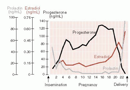
Biologic endocrinologists have spent considerable time and energy trying to tease out the maternal molecules. Although they have gotten close, there is still no definitive concoction of hormones that will immediately trigger maternal behavior in a nulliparous (virgin) rat. The leading culprits are estrogen, progesterone, and prolactin. An important ingredient appears to be the changing levels of the hormones. In Figure 17.2, note how the progesterone is seen to drop while the estrogen and prolactin rise in a rat just before delivery.
Oxytocin also plays some important role. Traditionally, we conceptualize oxytocin as the neuropeptide released from the posterior pituitary into general circulation, which leads to uterine contractions and milk ejection (see Figure 7.4). Recent research has found receptors for oxytocin within the brain, establishing central actions for this neuropeptide. Indeed, injecting oxytocin directly into the lateral ventricles in a rat will induce maternal behavior in a hormone-primed virgin rat. More recently, it has been shown that oxytocin levels in the paraventricular nucleus (PVN) increase with maternal aggression. Likewise, infusion of synthetic oxytocin into the PVN also increases maternal aggression toward an intruder.
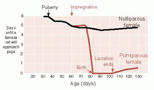
Further complicating this picture is the fact that hormones facilitate maternal behavior but are not required for it. Nulliparous rats will initially avoid new pups placed in their cage. However, if exposed over a series of days (1 hour each day), they will respond maternally to the pups within 5 to 6 days. Pregnant rats will show similar avoidance until after they have delivered. Then they will quickly display maternal behavior to any pup for the rest of their lives, proving that “once a mother, always a mother” (Figure 17.3).
The Brain
Taken together, these studies suggest that the hormonal fluctuations late in pregnancy act on the brain to decrease fear or aversion and increase attraction toward infants. Because maternal behavior persists once it is established, it is likely that the experience permanently changes some regions in the brain. An area that has been intensely studied is the one that we discussed in Chapter 16: the preoptic area (POA) located in the anterior hypothalamus.
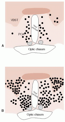
The POA is rich in estrogen, progesterone, prolactin, and oxytocin receptors, all of which increase during gestation. Lesions of the POA will disrupt maternal behavior. The POA appears to be a region that receives olfactory and somatosensory input and has projections to midbrain and brain stem nuclei. Numan and Sheehan describe an elegant experiment that demonstrates the central role of the POA with maternal behavior. Postpartum rats were exposed to either pups or candy for 2-hours. Then their brains were analyzed for the presence
of the transcription factor Fos. (Fos is used as a general marker of gene expression.)
of the transcription factor Fos. (Fos is used as a general marker of gene expression.)
Figure 17.4 shows the results of the study. This is a slice through the forebrain that includes the anterior hypothalamus on either side of the ventricle. (For a human comparison, see Figure 16.10.) Note the increased activation in the POA as well as other regions of the rat exhibiting maternal behavior.
Dopamine
Up to this point, we have stressed the importance of gonadal steroids and neuropeptides in the development of maternal behavior, but the neurotransmitter dopamine also appears to play an important role. We discussed in Chapter 12 the activation of the orbitofrontal cortex (OFC) and the ventral tegmental/nucleus accumbens area (see Figure 12.3) in the experience of pleasure. As might be expected, these areas are active in mothers.
One study scanned new mothers while they were looking at pictures of their own child and pictures of unfamiliar children. The mothers showed greater activation of the OFC when viewing their own child. With rats, researchers have found that mother rats will press a bar for access to pups the way they will press a bar for amphetamines or electrical stimulation. Additionally, pup exposure increases the release of dopamine at the nucleus accumbens. Alternatively, dopamine blockers will impair maternal behavior (see box). These studies give some neurobiologic explanations for the “joys of motherhood.”
Licking and grooming
This brings us to a series of studies from Michael Meaney’s laboratory in McGill University, which may be some of the most important recent neuroscience studies for mental health professionals. These studies tie together maternal behavior with lasting effects on the offspring’s behavior, hypothalamicpituitary-adrenal (HPA) axis, and even their DNA.
The story starts in the 1960s when researchers noted that pups “handled” once a day during the first weeks of life showed a reduced adrenocorticotropic hormone and corticosterone response to stress. Later, it was established that it was not the “handling” per se that produced this effect, but the mother’s increased licking of the pups when they
were returned to the nest. The mothers were simply trying to get the human odor off their pups and this extra attention to the pups resulted in their improved response to stress when they grew up to be adults.
were returned to the nest. The mothers were simply trying to get the human odor off their pups and this extra attention to the pups resulted in their improved response to stress when they grew up to be adults.
SCHIZOPHRENIA AND DOPAMINE BLOCKERS
Mothers who suffer from schizophrenia are known to be less involved with their children. They are generally more remote and less responsive during mother-infant play. This could be another example of the negative symptoms of the disorder. Worse yet, the problem might be exacerbated by the medications used to treat the patients.
In a recent study, Li et al. looked at the effect of injections of haloperidol, risperidone, and quetiapine on maternal behavior in rats. The antipsychotic medications inhibited maternal behaviors, such as nest building, pup licking, and pup retrieval. The figure shows the results for pup retrieval. Shortly after the injections, mothers failed to retrieve their own pups. Such studies suggest caution when treating human mothers with antipsychotic agents.
Meaney discovered naturally occurring strains of rats that licked and groomed their pups at different rates. This particular behavior occurs when the mother rat enters the nest and gathers her pups around her for nursing. She will intermittently lick and groom the pups as they nurse. Meaney named one group the high lick and groom (high L and G) mothers and the other the low lick and groom (low L and G) mothers.
In a flurry of experiments in Meaney’s laboratory, it was established that high L and G mothers produced offspring with subtle but significantly different brains. After 20 minutes of restraint (very stressful for a rodent), the rats from high L and G mothers secrete less corticosterone (Figure 17.5A). They also produce less corticotropin-releasing hormone messenger RNA (CRH-mRNA) in the hypothalamus (Figure 17.5B). Additionally, the amount of maternal licking and grooming correlates with the number of glucocorticoid receptors (GRs) in the hippocampus (Figure 17.5C). They literally produce more GRs as a result of enhanced gene expression.
In summary, a mother’s increased attention enhances the sensitivity of the HPA axis most likely by turning on the appropriate DNA. Offspring of attentive high-licking mothers demonstrate greater feedback to the hypothalamus by way of the increased GRs, which inhibits CRH production and corticosterone release. Perhaps most significant, the pups from a high L and G mother show a greater willingness to explore novel environments as adults and demonstrate enhanced resilience under duress.
Trading Places
In a follow-up study, Meaney et al. switched some of the mothers and pups. That is, pups from high L and G mothers were raised by low L and G mothers and vice versa. The results were stunning and show how behaviors and patterns emerge from combinations of genetic predisposition and environment. Figure 17.6 shows the behavior of the adopted female rats raised by high L and G mothers once they matured. They were more inclined to explore an open area and provided greater licking and grooming to their own pups. Note how the determining factor is not the genetic makeup, but the nurturing behavior of the mother that raised them. In other words, a low L and G female will become a high L and G mother if she is raised by a high L and G mother. So the behavior can be passed from generation to generation, but it is not genetic—it is epigenetic.
Effect on the DNA
Meaney and his group have taken this line of research to the next level by searching for epigenetic mechanisms that can explain the enduring effects the mother’s behavior has on the pups. Briefly, epigenetic molecular attachments on the DNA (see Figures 6.8 and 6.9) affect gene expression, which in turn alters the proteins produced—in this case the GR.
Meaney’s group identified a section of the rat DNA that encodes for hippocampal GR. Looking specifically at the promoter region of this DNA—the region where the transcription starts—they analyzed methylation of the cytosine-guanine sites (CG sites).
They found a much greater frequency of methylation for the low L and G group compared with the high L and G group along the GR promoter gene (Figure 17.7). In other words, the mother’s attentive behavior reduced the methylation of the gene and allowed for greater production of the GR—which in turn made the rats more resilient. This is an excellent example of the neuroscience model (see Figure 6.13) showing how environment, gene expression, and brain development affect behavior.
They found a much greater frequency of methylation for the low L and G group compared with the high L and G group along the GR promoter gene (Figure 17.7). In other words, the mother’s attentive behavior reduced the methylation of the gene and allowed for greater production of the GR—which in turn made the rats more resilient. This is an excellent example of the neuroscience model (see Figure 6.13) showing how environment, gene expression, and brain development affect behavior.
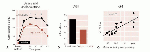 FIGURE 17.5 • Rats raised by a mother with a high frequency of licking and grooming behavior show a more modest corticosterone release in response to stress (A), less corticotropin-releasing hormone messenger RNA (CRH-mRNA) (B), and greater glucocorticoid receptors (GR) in the hippocampus. C. The correlation between GRs and licking and grooming by the mother. (Adapted from Liu D, Diorio J, Tannenbaum B, et al. Maternal care, hippocampal glucocorticoid receptors, and hypothalamic-pituitary-adrenal responses to stress. Science. 1997;277(5332):1659-1662.)
Stay updated, free articles. Join our Telegram channel
Full access? Get Clinical Tree
 Get Clinical Tree app for offline access

|
