Attention
INTRODUCTION
The last aspect of cognition that we will review is the ability to attend to external or internal stimuli. Like memory, attention is one of the oldest and most studied areas of cognitive science. One hundred years ago researchers used stopwatches and psychological tests to measure attention. In the latter half of the 20th century, the placement of microelectrodes into the brains of monkeys opened the door to investigating the capacity of individual neurons to attend. Now with brain imaging studies we can observe, in real time, the shifting focus of an awake human solving a puzzle—with or without medication. One hundred years of research has given us a better understanding of the power of the brain to focus on relevant stimuli, although many aspects of the neurobiology of attention remain mysterious.
Attention in a broad sense describes the mechanism that weighs the importance of various stimuli and selects the one that will receive the brain’s focus. The brain has limited capacity for attention. Numerous psychological tests have demonstrated the brain’s finite capacity to attend as more and more stimuli are added. A relevant example from modern life involves driving while talking on a cell phone. A recent study found that drivers using a cell phone had a fourfold increase in the chance of a serious accident. Handsfree phones were equally problematic. Attending to a conversation detracts the driver from attending to events on the road— contrary to what the driver thinks.
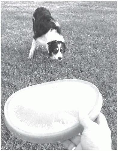
The capacity to concentrate and maintain one’s attention is inversely related to the ability to ignore other stimuli. Responding to other stimuli—whether internal or external—changes the brain’s focus. The brain cannot attend if it is wandering from one thought to another. The border collie in Figure 20.1 shows an example of highly selective attention. The dog is not only focused on the frisbee but also actively ignoring other objects of potential interest around him. In this state he will ignore female dogs, squirrels, children, and even food. He will not sniff the scent of other dogs or leave his own mark on the shrubbery. He appears to focus solely on the flying sphere.
Athletes provide another example of the intimate relationship between attending and ignoring. The quarterback who has dropped into the pocket and is able to ignore all the noise and violence around him while he searches for an open receiver is an extraordinary example of attending and ignoring.
Likewise, when the artist or the absent-minded professor gets lost in work, they are ignoring most sensory inputs coming into their brain. Treatments that improve attention may be effective because they improve the brain’s capacity to screen out the unessential.
Likewise, when the artist or the absent-minded professor gets lost in work, they are ignoring most sensory inputs coming into their brain. Treatments that improve attention may be effective because they improve the brain’s capacity to screen out the unessential.
Measuring Attention
Continuous performance tasks (CPTs) give an objective estimate of an individual’s attention and impulsivity. Subjects watch a computer screen and hit a button or click a mouse whenever a specified sequence of symbols or letters appears (Figure 20.2A). Such tests reflect the subject’s capacity to attend as well as the ability to restrain impulsive answers. Results are compared with normative data for individuals in one’s age group.
Attention changes across the life span. Figure 20.2B shows the percent errors on one CPT (Test of Variables of Attention) for 1,590 individuals from the ages of 4 to 80+. Note how attention improves with age until the senior years. These findings appear to roughly correlate with the myelination of the white matter in the frontal lobes (see Figure 8.8).
WORKING MEMORY
Prefrontal Cortex
Working memory describes what is actively being considered at any moment. If the conscious brain is actually a tiny person inside a control room orchestrating the body’s responses, working memory would be what he sees on the monitors in front of him. It is temporary, limited in capacity, and must be continually refreshed. Traditionally, working memory has been associated with the prefrontal cortex (PFC). It most certainly resides there, but may also include connections with the parietal lobes.
ADHD AND ADULTHOOD
Attention-deficit/hyperactivity disorder (ADHD) was once considered a childhood disorder. More recently, it has become popular for adults to get treatment for this condition. However, a recent meta-analysis of follow-up studies found that most children with ADHD fail to meet the full criteria for the disorder as adults—only 10% to 15%.
Trauma to the PFC impairs working memory. Phineas Gage is the most famous case that shows the effects of frontal brain damage on working memory. He was the young railroad worker who had a tamping iron explode through his frontal cortex (see Figure 14.4). He went from being responsible and organized to impulsive and inattentive. His inability to sustain a thought in his working memory could be considered the root cause of his wandering attention.
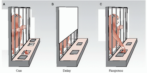
Researchers developed a way to test working memory in monkeys, called the delayed-response task. In this task, a monkey is shown a piece of fruit being placed in one of two randomly chosen receptacles (Figure 20.3). This is called the cue. A screen is then pulled down obscuring the monkey’s view and lids are placed over both receptacles. When the screen is lifted, the monkey gets one chance to remove the correct lid and receive his reward. The significance of this test is that the monkey must hold the visual image of the location of the fruit in his working memory during the delay period. Healthy monkeys learn this task quickly. Monkeys with frontal lobe lesions perform poorly.
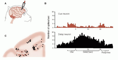
In the 1970s, researchers began putting microelectrodes into individual neurons in the PFC of monkeys while they participated in the delayedresponse task (Figure 20.4A). They found that neurons reacted differently during the task. Some neurons were active only during the cue and response periods, whereas other neurons became active during the delay period (Figure 20.4B).
The delay neurons start firing with the presentation of the cue and stop with the response. These neurons
seem to hold the memory of the task—literally the neural equivalent of working memory. When the monkeys incorrectly responded, the delay neurons were usually inactive. If the monkeys were distracted during the delay period, the delay neurons would usually settle down and the monkeys would make incorrect responses or not respond at all.
seem to hold the memory of the task—literally the neural equivalent of working memory. When the monkeys incorrectly responded, the delay neurons were usually inactive. If the monkeys were distracted during the delay period, the delay neurons would usually settle down and the monkeys would make incorrect responses or not respond at all.
After the studies, the researchers sacrificed the monkeys and identified the location of the microelectrodes. The results from several of the monkeys from one section of their right PFC are condensed and are shown in Figure 20.4C. The cue neurons and delay neurons are shown in different colors. Note how the delay neurons are more common than the cue neurons and cluster together. It is likely the delay neurons do not work independently but are part of a network that holds the image in working memory.
Catecholamines
Working memory is modulated by the catecholamines: dopamine (DA) and norepinephrine (NE). Pharmacologic interventions that increase DA and NE in the PFC enhance working memory and improve attention. Alternatively, agents that block DA receptors, such as haloperidol, have been shown to degrade performance on delayed-response tasks.
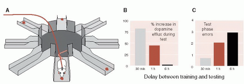
Phillips et al. examined the relationship between accuracy on a delayed-response task and DA release in the PFC. They placed microdialysis probes in the PFC of rats (see Figure 12.4A), which allowed continual analysis of extracellular DA concentrations while the rat performed the task. The rats were tested using an eight-arm radial maze (Figure 20.5A). In the training phase, rats were given 5 minutes to explore a radial arm maze that had four randomly chosen arms baited with food. During the delay, the rats were confined at the center of the maze in the dark from 30 minutes to 6-hours. In the test phase, food was placed on the opposite arms from the training phase and the rats were given 5 minutes to locate the rewards. Errors were scored as entries into unbaited arms. Extracellular DA was analyzed at baseline and during the testing phase.
EXECUTIVE FUNCTION VERSUS WORKING MEMORY
The terms executive function and working memory are often used synonymously in the literature. Some researchers prefer one term while others prefer the other, but they are not synonymous. Executive function includes working memory as well as other higher level cognitive skills such as organizing priorities and planning initiation strategies. Although executive function and working memory describe different functions in the brain, they share the same underlying mechanisms. Both reside in the PFC, are impaired with frontal lobe damage, and fluctuate with catecholamine modulation.
The results show an inverse correlation between extracellular DA in the PFC during the testing phase and errors (Figure 20.5B). When the delay was only 30 minutes, the efflux of DA into the PFC increased over the baseline by 75% and the rats made only few error journeys into unbaited arms. However, as the delay was increased, the DA efflux decreased and the errors mounted.
Biofeedback
Neurofeedback (also called electro encephalogram [EEG] biofeedback) is a treatment option that
allows patients to exercise their brain to improve attention and concentration. The EEG frequencies are divided into four groups (see Figure 15.3). β waves are the frequency pattern produced when a person is alert and concentrating. The goal of neurofeedback is for the patient to generate more β rhythm and less α and θ rhythms.
allows patients to exercise their brain to improve attention and concentration. The EEG frequencies are divided into four groups (see Figure 15.3). β waves are the frequency pattern produced when a person is alert and concentrating. The goal of neurofeedback is for the patient to generate more β rhythm and less α and θ rhythms.
 FIGURE 20.5 • A. Radial arm maze with four arms baited. B. Percent increase in extracellular dopamine at the testing phase. C. Errors at the testing phase. (Adapted from Phillips AG, Ahn S, Floresco SB. Magnitude of dopamine release in medial prefrontal cortex predicts accuracy of memory on a delayed response task. J Neurosci. 2004;24(2):547-553.)
Stay updated, free articles. Join our Telegram channel
Full access? Get Clinical Tree
 Get Clinical Tree app for offline access

|


