Fig. 1
T2-weighted MRI images (a) and lateral ventricular volume measurements (b) from rats 24 h after injection of 50 μl of saline or thrombin (3 U) into the right lateral ventricle. Values are means ± SD, n = 6, # p < 0.01 vs saline group
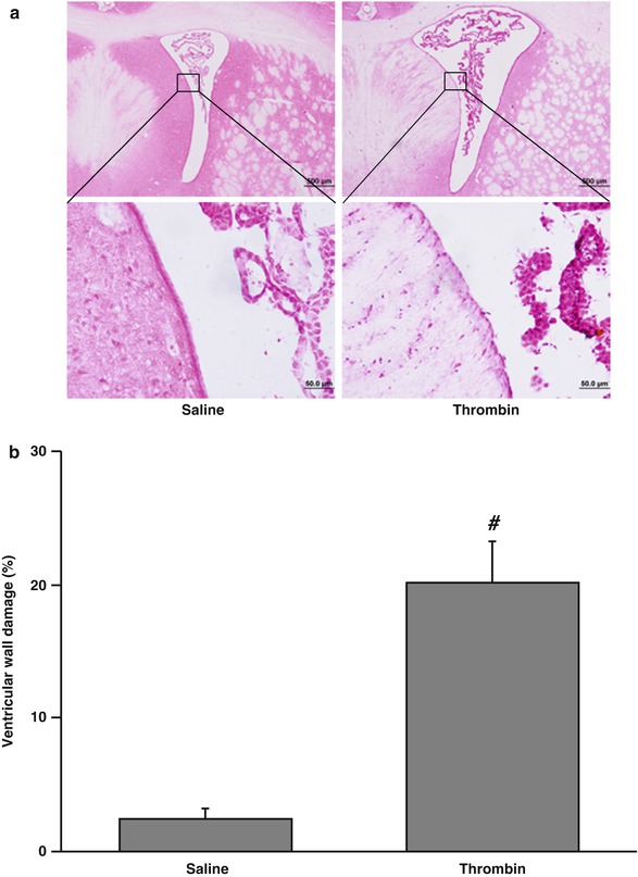
Fig. 2
The damaged ventricular wall in H&E-stained coronal sections (a) and the percentage of damage ventricular out of total ventricular wall (b) at 24 h after injection of 50 μl of saline or thrombin (3 U) into the right lateral ventricle. Values are means ± SD, n = 6, # p < 0.01 vs saline group
Systemic treatment with acetazolamide, a carbonic anhydrase inhibitor, 1 h after intraventricular injection of 3 U of thrombin resulted in less ventricular enlargement compared with vehicle treatment (16.1 ± 4.2 vs 29.5 ± 5.3 mm3 in the vehicle-treated group, p < 0.01) (Fig. 3).
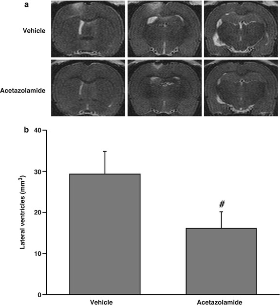
Fig. 3
T2-weighted MRI images (a) and lateral ventricular volumes (b) 24 h after injection of thrombin (3 U). Rats were treated systemically (IP) with vehicle or acetazolamide 1 h after the thrombin was injected. Values are means ± SD, n = 6, # p < 0.01 vs vehicle-treated group
Discussion
In this study, we found intraventricular injection of thrombin caused hydrocephalus and ventricular ependymal wall damage. Acetazolamide, given at 30 mg/kg IP 1 h after thrombin injection, attenuated thrombin-induced hydrocephalus.
Thrombin is a serine protease and an essential component in the coagulation cascade. Experimental investigations have indicated that thrombin formation plays a major role in ICH-induced injury [9, 11, 16]. Thrombin is responsible for early brain edema formation after ICH, and that edema results partly from a direct opening of the blood-brain barrier (BBB). We have demonstrated that intraventricular injection of thrombin can cause significant ventricular dilatation and periventricular parenchyma injury [6]. At present, the mechanisms associated with thrombin-induced ventricular dilatation are unknown. Ependymal injury such as reduced cilia and abnormal of organelle structure were observed in thrombin-injected rats [6]. Ependymal damage may lead to increased periventricular brain injury and to hydrocephalus [4, 14]. Activities of normal ependymal cilia are thought to direct cerebrospinal fluid (CSF) current toward the ventricular outlets. Previous study showed absent or functionally defective ependymal cell motile cilia may be a cause of hydrocephalus in a mouse model [1]. Therefore, ependymal damage and abnormal ependymal cilia may play a role in hydrocephalus induced by thrombin.
Acetazolamide, a carbonic anhydrase inhibitor, decreases CSF production in animal models [5, 10, 17]. Acetazolamide has a rapid onset of action. Treatment with 10 mg/kg of acetazolamide resulted in a significant decrease in CSF production and absorption within 3 h of ingestion in dogs [17]. Acetazolamide is also used in both children and adults to treat hydrocephalus and pseudotumor cerebri [6, 7]. We have shown that co-injection of acetazolamide reduced ICH-induced brain injury [7]. In the present study, acetazolamide treatment significantly reduced thrombin-induced hydrocephalus, most likely by reducing CSF production.
Conclusions
Intraventricular injection of thrombin caused ventricular wall damage and hydrocephalus in rats. Inhibition of carbonic anhydrase by acetazolamide effectively reduced thrombin-induced hydrocephalus. The results suggest that thrombin-induced hydrocephalus may result from reduced absorption and/or increased production of CSF.
Acknowledgment
Related posts:
 of Behavioral Deficits in Rodents Following Brain Injury Across Species, Gender, and Experimental Model
 Infarction After Aneurysmal Subarachnoid Hemorrhage
 Volume Determination in Subarachnoid Hemorrhage Using Rats
 Endothelial Growth Factor in Brain Edema Formation After Subarachnoid Hemorrhage
 in Brain Swelling and Infarction Volume over Four Days After Hypoxia Ischemia in Neonatal Rats
 of Gender on Iron-induced Brain Injury in Low Aerobic Capacity Rats
Stay updated, free articles. Join our Telegram channel

Full access? Get Clinical Tree


