Avoiding Complications and Correcting Errors in Movement Disorder Surgery

14 Avoiding Complications and Correcting Errors


Philip A. Starr


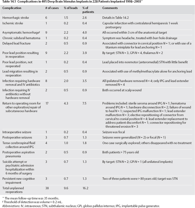
Several recent publications have reviewed complications of deep brain stimulation (DBS)18 and lesioning surgery4,912 for movement disorders. A summary of our group’s perioperative and device-related complications in 405 DBS implants for movement disorders is provided in Table 14.1. Procedures were performed with frame-based stereotaxy using magnetic resonance imaging (MRI) and microelectrode recording (MER). Medtronic Activa (Medtronic, Inc., Minneapolis, MN) DBS hardware was used in all cases. The total incidence of unexpected returns to the operating room for management of a complication was 38 cases, or 9.6% of implanted leads.


This chapter describes our current methods for complication avoidance and management. These methods continue to evolve. Although this discussion is oriented primarily toward DBS, many of the principles apply to other stereotactic procedures. Complications uniquely associated with stereotactic lesioning procedures are discussed briefly at the end of the chapter.


Operative Complications


Stroke


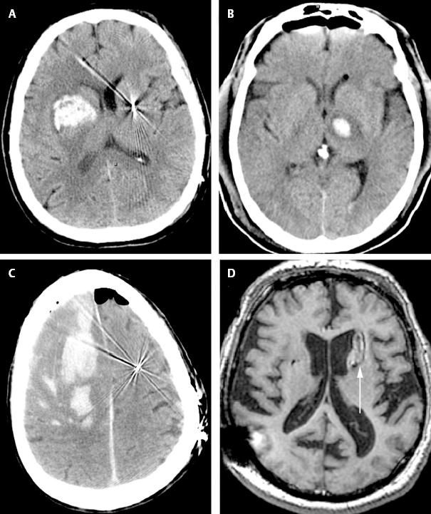
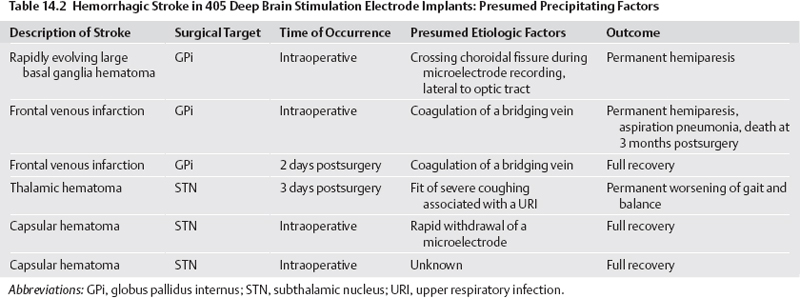
Stroke is the most serious potential complication of movement disorders surgery. Stroke is defined as a new neurological deficit of vascular origin, lasting longer than 24 hours. Using this definition, our DBS series includes seven strokes, for an incidence of 1.7% per lead and 3.0% per patient. Six of these were hemorrhagic strokes. Fig. 14.1 shows postoperative brain imaging for representative hematomas. Table 14.2 provides details of presumed etiologic factors.


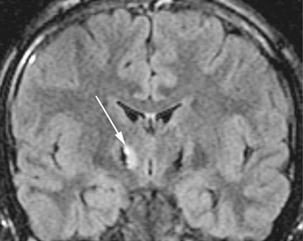
Ischemic infarction is very infrequent following DBS surgery, occurring only once in our series (Fig. 14.2). In contrast, delayed ischemic capsular infarction has been well described following pallidotomy10,13 and is probably a more frequent complication of stereotactic lesioning surgery than of DBS.


Asymptomatic hemorrhage is more common than symptomatic hemorrhage but is only detected if postoperative imaging is performed systematically. Our rate of asymptomatic hemorrhage, detected on routine postoperative magnetic resonance imaging (MRI) was 2.2% per lead. A typical asymptomatic hematoma is shown in Fig. 14.1D. Most of the asymptomatic hematomas occurred subcortically, 25 to 35 mm superior to the target, corresponding to the location where the guide tube for the microelectrode terminated.


Avoidance of Stroke

Based on our experience, we recommend the following measures to reduce the incidence of stroke:


  1. Maintain systolic pressure under 140 and mean arterial pressure (MAP) under 90. In any patient with preexisting hypertension, we place an arterial line and control blood pressure with a continuous intravenous drip (esmolol, nitroglycerine, or nitroprusside).
  2. Avoid damage to, or coagulation of, venous structures. We perform surgeries through burr holes where the surface vessels may be directly visualized, rather than a twist drill hole. In addition, subsequent to the two venous infarctions in our series, our stereotactic MRI protocol has included a contrast-enhanced T1-weighted image set to visualize cortical veins as well as arteries. Surgical planning software is then used to plan a trajectory to the target that avoids MRI-visible blood vessels, sulci, and the ventricles.
  3. During pallidal surgery, avoid passing instruments deep to the optic tract (OT), into the choroidal fissure. One of our worst DBS complications (Fig. 14.1A) was associated with bleeding from a presumed choroidal vessel as we attempted to localize the optic tract. Currently, if we do not locate OT within 2 mm of the base of pallidum, we do not continue more inferiorly.
  4. Any sudden Valsalva during or immediately after surgery carries a risk of hemorrhage. If a patient has an upper respiratory infection or any other reversible source of coughing, surgery should be postponed. Smokers with a chronic cough must be advised that their risk of perioperative hemorrhage may be increased.
  5. Avoid rapid insertion or withdrawal (> 0.5 mm/s) of instruments into or out of the brain.
  6. During and at the end of the procedure, cover the cortical entry with Gelfoam (Pfizer Inc., New York, NY) and fibrin glue, to avoid subdural blood accumulation.

Management of Stroke

Should a new neurological deficit occur during the procedure, surgery is immediately stopped. The anesthesiologists are asked to redouble efforts at blood pressure control and ensure appropriate oxygenation. All instruments are removed from the brain and the cortex is inspected. Superfcial bleeding sources are coagulated and surgery continues. If no surface bleeding is seen and no bleeding is seen from the cortical entry, the scalp is rapidly closed, the headframe is removed, and the patient is taken for a computed tomographic (CT) scan.


image

Short of a neurological deficit, there may be other more subtle signs of intraparenchymal hemorrhage that is initially asymptomatic and may remain so depending on its final size and location. When we have observed blood coming from the subcortical guide tube after withdrawal of a stylet or microelectrode, a small hematoma is almost always observed on postoperative imaging at a depth corresponding to the termination of the guide tube. During MER,if a significant region of electrical silence is observed at a depth where neuronal tissue is predicted based on prior adjacent MER tracks, the cause may be a small hematoma. If either of these signs of potential hematoma formation is observed, we halt the procedure and closely observe the patient for subtle new neurological deficit while recon-firming strict blood pressure control. If no deficit occurs in 5 to 10 minutes we proceed with surgery.


image

Fig. 14.1 Postoperative axial computed tomographic (CT) scans (A–C) or magnetic resonance imaging (MRI) (D) of hemorrhages complicating stereotactic deep brain stimulation (DBS) surgery in our series. (A) Hematoma that occurred during penetration of a mi-croelectrode into the choroidal fissure, lateral to the optic tract, during microelectrode mapping of globus pallidus internus (GPi). (B) Hematoma that occurred during rapid withdrawal (> 1 mm/s) from the subthalamic nucleus (STN). (C) Hemorrhagic venous infarct that occurred following inadvertent interruption and coagulation of a bridging dural vein. (D) An asymptomatic hematoma in the left caudate nucleus (white arrow), immediately anterior to the DBS lead. (The hyperintensity in the right parietal cortex is artifact from the overlying connector of the lead extender).


image

image

Fig. 14.2 Coronal magnetic resonance imaging (MRI) (FLAIR sequence) showing a delayed ischemic capsular infarction that occurred 1 week after bilateral subthalamic nucleus deep brain stimulation (DBS). The infarcted area (white arrow) is adjacent to the right DBS lead.


Infection


In our series, the incidence of serious infection, defined as infection requiring a return to the operating room for removal of all or part of the DBS hardware, was eight cases, or 2.0% per lead and 3.5% per patient (Table 14.1). All of these infections have occurred subcutaneously, starting at the lead extender or the implantable pulse generator (IPG). The major offending organisms were Staphylococcus (staph) aureus and Staph epidermidis. We have had no infections in the brain, and in fact cerebral abscess or cerebritis complicating DBS has not been reported in most recent series. Most of our infections presented with some combination of swelling, redness, pain, or drainage over the connector of the lead extender, or over the IPG. Most presented within 1 to 8 weeks of surgery, although one presented as a stitch abscess over an anchoring suture on the connector 2 years after implantation.


Infection Avoidance

Our approach to avoidance of infection, in addition to the obvious meticulous attention to sterile technique, is as follows:


  1. Preoperative prophylaxis with an antistaphylococcal cephalosporin at least 30 minutes prior to skin incision, followed by a second dose given 3 hours later, and three postoperative doses.
  2. Utilization of the lowest-profile hardware available, with recessing of larger components into a drilled bone trough in patients with thin skin.
  3. Emphasis on performing surgery efficiently to reduce operative time.
  4. Copious irrigation of all incisions with bacitracin solution prior to closure.

Infection Management

The management of hardware infections has not been standardized. In the report of Oh et al,6 infections of any part of the device were ultimately treated with removal of all hardware, despite initial attempts at more localized treatment. Our approach is as follows:


  1. For superficial infections at incision sites where hardware does not appear to be in direct contact with pus or necrotic tissue, the patient is treated with antibiotics and local wound care without hardware removal, followed weekly with clinical examination until wounds are completely healed.
  2. For infections where the lead extender or IPG is in direct contact with pus or necrotic tissue, the affected components are removed immediately on presentation, and then the patient is treated with the appropriate intravenous (IV) antibiotics. If a localized infection around the pulse generator or lead extender is discovered early in its course, this strategy may result in salvage of the brain electrode.
  3. An infection in direct contact with the brain lead or an infection along the extender or IPG that has been incubating for many weeks will usually necessitate removal of the DBS lead as well as other involved hardware.
  4. In cases where lead removal was necessary, we have been able to reimplant another DBS lead 2 to 3 months after full wound healing, without further infectious sequelae.

Sterile Fluid Collections


Four of our patients have presented within 1 month post-surgery with tense swellings around the IPG, which were fluctuant but painless and without redness or warmth. Surgical exploration of one of these revealed a sterile clear fluid collection consistent with cerebrospinal fluid (CSF). This appears to occur more frequently with burr hole– based anchoring methods that are not watertight (such as the Stimloc system from Medtronic, Inc., www.medtronic.com/physician/activa) because CSF can track down the hardware to accumulate in the pectoral cavity. Sealing the burr hole with Gelfoam and fibrin glue prior to closure has reduced the incidence of these sterile collections. If a swelling around an IPG is not red, tender, or warm, and the incision is healed, our practice is to observe it. Sterile fluid collections typically resolve spontaneously.


Editor’s Comments


Prevention of complications starts with the initial patient evaluation (Chapters 4 and 5) and continues through to intraoperative technique (Chapters 7 through 12). Nevertheless, complications will occur. The most common and most severe operative complication is that of hemorrhage. Subdural hematomas are uncommon but can occur in any intracranial procedure. To avoid this complication, anything that can raise intracranial pressure and tear diploic veins such as coughing or bucking must be avoided. In addition, probes should enter through a gyrus in which the pia-arachnoid has been opened to prevent distortion of the cortex and potential tearing of intracranial vessels. This is especially true of the DBS lead, and we recommend direct visualization of the probe into the brain. Operations close to the sinus can result in injury to the sinus or major bridging veins resulting in venous infarction. Scanning to the top of the head, using contrast to visualize these vessels directly, and placing the burr hole in a position that would not put them at risk can minimize the risk of this complication. Although rectilinear approaches are essential for mapping, a slight lateral to medial approach of 1 to 4 degrees is insignificant in its distortion of the map but may provide additional distance from the midline to enhance safety. The more common and more severe problems occur with intraparenchymal hemorrhages. The StealthStation (Medtronic Navigation, Louisville, CO) is especially useful in planning trajectories that will avoid not only cortical veins but also deep sulci that may curve back into the trajectory or deep venous structures such as the veins within the lateral ventricle. Slow, smooth descent of the probe is essential to minimize trauma. Absolutely critical is control of blood pressure during these procedures because even a normally hypotensive patient can become hypertensive during the stress of awake surgery.


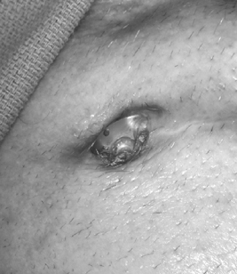
The most common immediately postoperative problem is that of infection. “Pickers” cannot leave the wound alone and will consciously or unconsciously pick at it. This behavior puts the implant at risk of infection. Pickers are frequently children and cognitively impaired patients and use of subcuticular sutures combined with tissue adhesive and careful monitoring can help decrease the risk. Beware of a history of self-mutilation, obsessive compulsion, and psychosis. We use antibiotics before, during, and after surgery in an attempt to keep the infection rate to an absolute minimum. This aggressive approach has resulted in a very low infection rate of 2%. Intraoperative antibiotics are generally vancomycin for its intracranial penetration and all instruments that enter the brain are wiped down with antibiotic-containing solution. Postoperatively, the IV antibiotics are used as long as the patient is in the hospital. On discharge, the patients are converted to oral antibiotics, and cephalosporins are generally used because the main concern is extracranial staph infections. Infections usually occur in a regional manner and can be treated as such. We have had to remove only one entire DBS system. Infections at the burr hole can be minimized using surgical techniques as described in Chapter 7. Since using low-profile caps and pericranial coverage in over 200 DBS lead placements, we have not had a single infection at the burr hole cover. The most common place where we encounter infections at this time is at the connector. The skin here is relatively thin and the device relatively large. These patients frequently have head trauma, and any laceration should be treated aggressively with antibiotic coverage if it is anywhere near the lead, connector, or extension wire. The use of lower-profile connectors, placing the connector above the incision, and covering the extension lead with an additional layer of temporalis fascia will, it is hoped, reduce this complication. It should also be emphasized to the patient to keep pressure off of this area whether from caps, wigs, or lying on hard surfaces in an attempt to encourage wound healing and diminish tissue necrosis. Operative removal and antibiotic treatment of erosions are the same as for overt infections. Any skin erosion should be treated as an infection even if the skin appears intact over it (Fig. 14.3).


Stitch abscesses and any potential superficial infections in the scalp are aggressively treated with antibiotics. Failure to resolve this after a course of antibiotics requires intraoperative evaluation, de-bridement, and removal of the contaminated equipment, while attempting to preserve the lead. The burr hole cover usually requires placement with a metal plate to ensure security of the lead while removing as much of the contaminated plastic as possible. Long-term IV antibiotic with staph coverage and the addition of rifampin, which frequently acts synergistically, has been successful in cleaning up these types of infections. In the area of the connector, the connector boot is removed and a new boot placed, if this is the only area that has been contaminated. If there is wider contamination, then the extension lead must be removed in addition. First disconnecting it in the chest and leaving IPG in the chest and then pulling the remaining wire to the contaminated surface perform this. The contaminated area is opened last to prevent cross-contamination of the chest area. Vigorous antibiotic irrigation and the use of dilute hydrogen peroxide are recommended. A new connector can be attached to the lead to facilitate its identification at a later time. This connection should be displaced away from the area that is infected. Again, long-term IV antibiotics are recommended using a cephalosporin and rifampin. Infection in the chest usually requires examination of the connector first, and if this area is not infected then the extension lead is cut below the connector. Attention is then drawn to the chest, which is opened, and the entire IPG and extension lead are then removed. Vigorous intraoperative antibiotic irrigation and postoperative IV antibiotics are similar to those described earlier, but the final choice is based on culture and sensitivity results. Drains are not necessary.


image

Only gold members can continue reading. Log In or Register to continue

Stay updated, free articles. Join our Telegram channel

Aug 5, 2016 | Posted by in NEUROSURGERY | Comments Off on Avoiding Complications and Correcting Errors in Movement Disorder Surgery

Full access? Get Clinical Tree

Get Clinical Tree app for offline access