, John A. Ameriks2, Frank T. Jordan3 and James M. Giuffre4
(1)
Center for Diseases and Surgery of the Spine, Las Vegas, NV, USA
(2)
Nevada Surgical Group, Las Vegas, NV, USA
(3)
North Vista Hospital, Las Vegas, NV, USA
(4)
International Spinal Development and Research Foundation, Las Vegas, NV, USA
1.
The balloon-assisted endoscopic retroperitoneal gasless (BERG) approach is executed with one spinal surgeon, one vascular/general surgeon, and one endoscopically trained technician (Fig. 32.1).
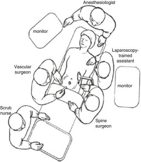

Fig. 32.1
Operating room setup and patient positioning
2.
The patient is placed in the supine position. Following general anesthesia, the patient is draped and prepped in standard fashion and preoperative antibiotics are given. Fluoroscopy is used to find the landmarks of the appropriate lumbar level. The skin is marked identifying the level and angle of the pathologic disc interspace(s). These marks are drawn on the lateral aspect of the left abdomen, marking the angles of the disc spaces to be addressed (Fig. 32.2).
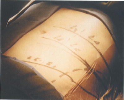

Fig. 32.2
Following fluoroscopy, the angle of disc spaces to be addressed are marked on the lateral aspect of the abdomen. The initial left flank incision is made approximately 1 cm above the iliac crest in the midaxillary line
3.
A transverse 20-mm left flank incision is made approximately 1 cm above the left iliac crest in the midaxillary line (Fig. 32.2). The dissection is taken down through the external oblique, internal oblique, and transversus muscles under direct vision to the preperitoneal fat layer using a clear-ended, laparoscopic dissecting port.
4.
The retroperitoneal space is then gently insufflated with a bulb syringe and then digitally dissected into the iliac fossa to allow for balloon insertion. An undeployed elliptical-shaped preperitoneal balloon (PDB-2; Origin Medsystems, Meno Park, CA) is advanced through the incision until the entire balloon is within the retroperitoneal space (Fig. 32.3).
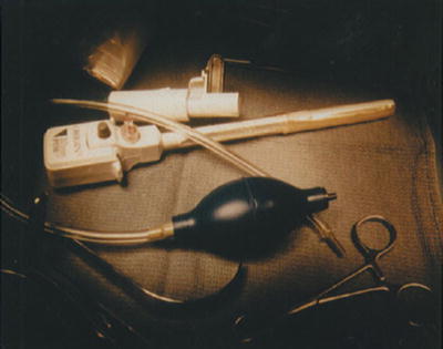

Fig. 32.3
The PDB-2 (preperitoneal dissection balloon-2)
5.
A 0-degree-angle endoscope is placed through the lumen of the dissection cannula, and the balloon is expanded to an approximate volume of 1 L (Fig. 32.4).
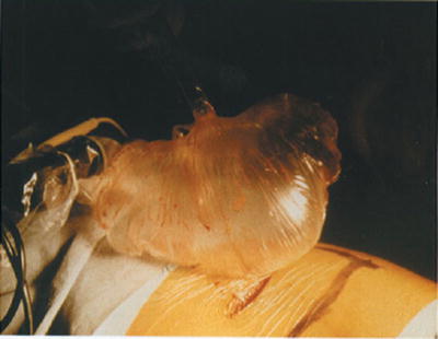

Fig. 32.4
The PDB-2 expanded to an approximate volume of 1 L
6.
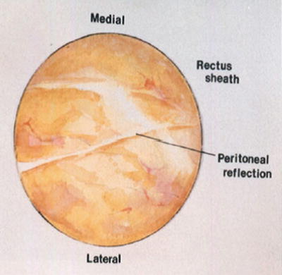
The endoscope is directed toward the anterior abdominal wall; this allows the identification of the peritoneal reflection on the anterior abdominal wall, at the rectus sheath above and below the line of Douglas (Fig. 32.5).

