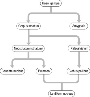
Basal ganglia
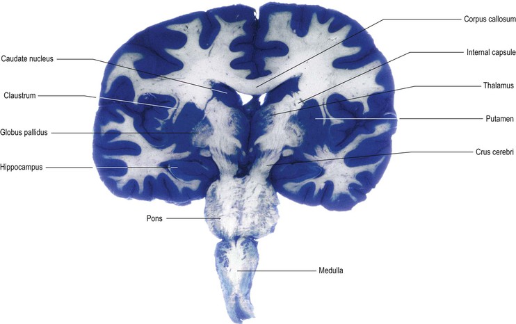
Within the cerebral hemisphere lie a number of nuclear masses. The most prominent of these are the caudate nucleus, putamen and globus pallidus which lie in close proximity to the internal capsule. They are collectively referred to as the basal ganglia, basal nuclei or corpus striatum (Figs 14.1, 14.2; see also Figs 13.3–13.9, 13.14). The amygdala, which is located within the temporal lobe, near to the temporal pole, has a similar embryological derivation but is functionally different, being part of the limbic system (Ch. 16). The most rostral and ventral part of the corpus striatum includes a region known as the nucleus accumbens (Fig. 13.6). This has connections similar to the rest of the corpus striatum and also has close connections with the amygdala, thus providing an important link between the basal ganglia and the limbic system.
The basal ganglia are primarily concerned with the control of movement. Disorders of the basal ganglia are manifest by abnormalities of motor control, posture and muscle tone. Their interface with the limbic system also confers a role in the physical expression of behaviour driven by affective and motivational states. The nucleus accumbens is associated with reward and gratification and is believed to be a part of the mechanism underlying addictive aspects of behaviour, including being an important site of action of addictive substances.
The caudate nucleus, putamen and globus pallidus are anatomically and functionally closely related to each other and are principally involved in motor control. The basal ganglia have important connections with other regions of the brain, particularly the cerebral cortex, the thalamus and subthalamic nucleus of the diencephalon, and the substantia nigra of the midbrain.
For gross anatomical reasons, the putamen and globus pallidus are sometimes together called the lentiform (or lenticular) nucleus (or complex). This is because they lie close together, forming an apparently single structure. The name means ‘lentil-shaped’, but a closer analogy is a Brazil nut or the segment of an orange. The lentiform nucleus is three-sided, having a convex lateral surface and two other surfaces that converge to a medial apex which lies against the genu of the internal capsule. The term lentiform, or lenticular, is rather archaic and of limited use, although it is still retained in certain anatomical names (such as the retrolenticular part of the internal capsule).
On phylogenetic, connectional and functional grounds, however, the putamen is more closely allied to the caudate nucleus than to the globus pallidus. The globus pallidus is, in phylogenetic terms, the oldest part of the corpus striatum and is sometimes referred to as the paleostriatum. The abbreviation pallidum is more commonly used, particularly in composite terms for afferent and efferent connections, such as subthalamopallidal or pallidothalamic.
The caudate nucleus and putamen constitute the phylogenetically more recent neostriatum and are best regarded as a single entity. The two parts are almost (but not entirely) separated by the anterior limb of the internal capsule but their gross anatomical separation is not as significant as their neuronal and functional similarities. Together, they are commonly referred to simply as the striatum.
These rather confusing relationships are summarised in Figure 14.3.
Topographical anatomy of the basal ganglia
Striatum
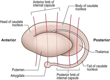
The striatum (neostriatum) consists of the caudate nucleus and the putamen. Their combined three-dimensional shape is reminiscent of a tadpole, when viewed laterally (Fig. 14.4).
Putamen
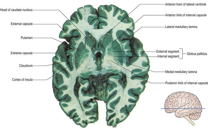
The putamen lies lateral to the internal capsule and globus pallidus (Figs 14.1, 14.2; see also Figs 13.7, 13.8, 13.14). It is separated from the globus pallidus by a thin lamina of nerve fibres, the lateral medullary lamina. Lateral to the putamen lies more white matter, sandwiched within which lies a thin sheet of grey matter, known as the claustrum. This separates the white matter into two layers, the external capsule and the extreme capsule (Fig. 14.1). Lateral to the extreme capsule lies the cortex of the insula, deep within the lateral fissure of the hemisphere.
Caudate nucleus
The caudate nucleus consists of a large head and a tapering, curved tail. The head of the caudate is almost completely separated from the putamen by the internal capsule. At its rostral extremity, however, it is continuous with the putamen through and beneath the anterior limb of the internal capsule (Fig. 14.4, see also Figs 13.4–13.6). At this anterior level, the most ventral and medial part of the striatum constitutes the nucleus accumbens. The head of the caudate nucleus forms a prominent bulge in the lateral wall of the anterior horn of the lateral ventricle (Fig. 14.1, see also Figs 13.3–13.7). The tail of the caudate passes posteriorly, gradually tapering as it does so and, following the curvature of the ventricle, descends into the temporal lobe where it lies in the roof of the inferior horn (Fig. 14.4, see also Figs 13.9–13.12).
Globus pallidus
The globus pallidus lies medial to the putamen, separated from it by the lateral medullary lamina. Its medial apex nestles into the lateral concavity of the internal capsule (Fig. 14.1). The globus pallidus consists of two divisions, referred to as the external (or lateral) and the internal (or medial) segments. These are separated by a thin sheet of fibres, the medial medullary lamina (Fig. 14.1). The smaller internal segment shares many similarities, in terms of cytology and connections, with the pars reticulata of the substantia nigra in the midbrain. Although the two are separated by the internal capsule, these are best regarded as a single entity, in the functional sense.
Substantia innominata
The term substantia innominata refers to the basal part of the rostral forebrain that lies beneath the corpus striatum (see Fig. 14.7). This complex region contains several groups of neurones, one of them being the nucleus basalis (of Meynert), that project widely to the cerebral cortex and utilise acetylcholine as their neurotransmitter. These neurones undergo degeneration in Alzheimer’s disease.
Functional anatomy of the basal ganglia
Connections of the striatum
The caudate nucleus and putamen, together commonly referred to as the striatum, are best considered as a single entity since they share common neuronal organisation, neurotransmitter systems and connections (Fig. 14.5). They are often regarded as the ‘input’ portions of the basal ganglia, since the majority of afferents from other parts of the brain end here rather than in the globus pallidus.

< div class='tao-gold-member'>
Related posts:

Stay updated, free articles. Join our Telegram channel

Full access? Get Clinical Tree


