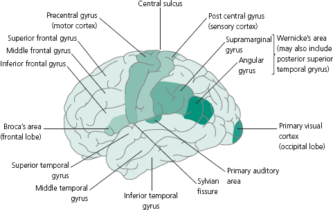
Cortical Anatomy
Figure 2.1 demonstrates the following relevant key areas on the cortical surface:
Remember: The association between dominant hemisphere and speech areas and therefore a further important point about ascertaining handedness of patients at the beginning of obtaining a neurological history.
Figure 2.1 Lateral surface of cerebral hemisphere demonstrating cortical surface anatomy.
- Primary motor cortex: Situated anterior to central sulcus (frontal lobe) in the precentral gyrus; involved in contralateral motor function—Brodmann’s (Br.) Area 4.
- Primary somatosensory cortex: Situated posterior to central sulcus (parietal lobe) in the postcentral gyrus; involved in contralateral sensory function—Br. Areas 1, 2 and 3.
- Motor speech area: In the dominant hemisphere only (left hemisphere for right-handed subjects and usually left hemisphere for left-handed people); anatomically situated in the inferior frontal gyrus (pars triangularis and opercularis); also known as ‘Broca’s area’ and involved in speech output—Br. Area 44.
- Wernicke’s area: In the dominant hemisphere; anatomically situated in the supramarginal gyrus—Br. Area 40 (part of inferior parietal lobule) and posterior part of superior temporal gyrus; involved in comprehension of speech.
- Primary visual cortex: In the occipital lobe adjacent to the calcarine sulcus—Br. Area 17; part of the visual pathway.
Brainstem and Cranial Nerves (CN)
The midbrain is the most rostral part of the brainstem; CN III (oculomotor) and IV (trochlear) arise from the midbrain. The pons is situated between the midbrain and the medulla with CN V (trigeminal), VI (abducens), VII (facial) and VIII (vestibulocochlear) nerve entering into or exiting from ventral pons. The medulla is caudal to pons being continuous with spinal cord. Fibres of the CN IX (glossopharyngeal), X (vagus) and XII (hypoglossal) nerves enter into or exit from the ventral aspect of the medulla. The basic clinical features of lesions associated with cranial nerves are presented in Table 3.3.
Only gold members can continue reading. Log In or Register to continue


