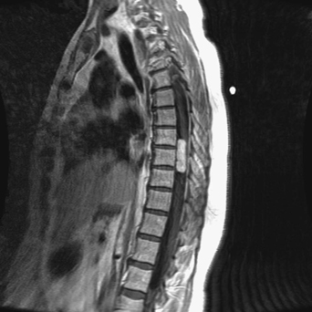
64 A 55-year-old woman had a year and a half of history of midthoracic pain with a sensory deficit and bowel and bladder dysfunction. Her symptoms worsened over time. Sagittal gadolinium magnetic resonance imaging (MRI) of the thoracic spine demonstrates a T8–T10 enhancing intradural extramedullary lesion (Fig. 64-1). FIGURE 64-1 Gadolinium-enhancing thoracic intradural extramedullary lesion as seen on thoracic MRI. Thoracic meningioma A thoracic laminectomy with microresection was done. Spinal meningiomas encompass 25 to 40% of extra-medullary primary spinal tumors, and are more prominent in middle-aged women. The mean time before diagnosis is about a year, and the most common complaint is paraparesis followed by radicular pain. A thoracic location is the most frequent, then cervical. An anterior or anterolateral location is slightly more common than a posterior or posterolateral location. The meningothelial subtype is the most popular. Surgical resection usually results in improvement. The calcified component of the meningioma and the ventral component (if present) are the riskiest aspects of resection. In addition, extradural spinal meningiomas, although rare, are higher risk tumors. They are believed to arise from the segment of the nerve root where the arachnoid is in contact with the dura. These occur in younger male patients, and can be more aggressive. A key element to a successful surgery is the use of microsurgical technique along with intraoperative neurophysiologic monitoring. Tumor resection should attempt devascularization along its dural attachment. Piecemeal resection is undertaken to avoid cord manipulation. The use of intraoperative ultrasound can optimize tumor localization. Klekamp J, Samii M. Surgical results for spinal meningiomas. Surg Neurol 1999;52:552–562
Benign Thoracic Tumor
Presentation
Radiologic Findings

Diagnosis
Treatment
Discussion
SUGGESTED READING
< div class='tao-gold-member'>
Benign Thoracic Tumor
Only gold members can continue reading. Log In or Register to continue

Full access? Get Clinical Tree


