Fig. 1
Brain water content and neurobehavior differences after SBI. (a) Brain water content (sham n = 7, SBI n = 12). (b) Composite Garcia neuroscore (sham n = 7, SBI n = 12). (c) Beam walking score (sham n = 7, SBI n = 12) *p < 0.05 vs Sham
Neurobehavior
Twenty-four hours after SBI, before euthanasia, animals were subjected to four neurobehavioral tests: composite Garcia neuroscore (neuroscore), beam walking test, corner turn test, and beam balance test.
Composite Garcia Neuroscore
The neuroscore of injured animals (15.8 ± 0.52) was statistically lower than that of sham animals (19.71 ± 0.18) (p < 0.05, Fig. 1b) 24 h after SBI. However, while statistically lower than SBI-injured animals, the neuroscore did not significantly correlate with the brain water content of the right-frontal lobe 24 h post-ictus (ρ = −0.02, p > 0.05, Fig. 2a).
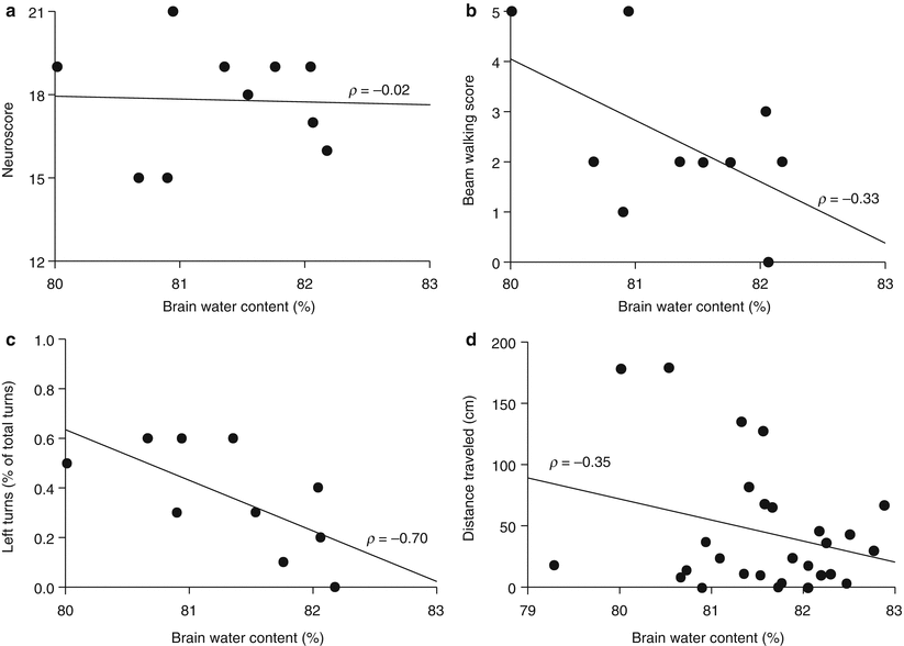
Fig. 2
Neurobehavior correlation with brain water content in animals subjected to SBI. (a) Composite Garcia neuroscore (n = 10). (b) Beam walking score (n = 10). (c) Corner turn test (n = 10). (d) Beam balance test (n = 35). All correlations were tested for correlation using Spearman’s rank correlation coefficient (ρ) and statistical significance (p-value)
Beam Walking
Corner Turn Test
The number of left turns in the corner turn test significantly correlated with the brain water content of the right-frontal lobe 24 h following SBI (ρ = −0.70, p < 0.05, Fig. 2c).
Beam Balance Test
The distance traveled for the beam balance test also significantly correlated with brain water content (ρ = −0.35, p < 0.05, Fig. 2d).
Discussion
SBI pathophysiology develops over several hours after the initial insult. The primary injury consists of the initial lobectomy and intraoperative hemorrhage [11]. Secondary injury occurs within hours of resection and includes brain edema, neuroinflammation, and postoperative hematoma [10, 11]. As with most neurological diseases, brain edema is a predictor of poor outcome in patients. Therefore, development of novel therapies for reducing or preventing cerebral edema is key to lowering the costs of SBI.
The rat model of SBI, developed by Vikram et al. [11], mimics the damage to healthy tissue that is unavoidable during many neurosurgical procedures. Post SBI, rats experience an increase of 4–5 % in brain water content of the right-frontal lobe, causing tissue swelling [11]. The current study utilized the rat SBI model to identify the appropriate sensorimotor tests for evaluating drug efficacy after SBI.
Herein, four common and inexpensive sensorimotor tests were examined for their ability to predict the magnitude of cerebral edema after SBI. The composite Garcia neuroscore is a combination of several subtests evaluating sensorimotor function [5, 13]. Furthermore, the composite Garcia neuroscore has the benefit of identifying deficits caused by unilateral injury because a number of the tests examine left versus right function, such as axial sensation and limb movement symmetry [5, 13]. Despite the fact that statistically significant functional deficits are found between SBI-injured animals and sham animals (Fig. 1b), they did not correlate with the brain water content. This is likely due, in part, to the inclusion of neuroscore subtests that do not identify unilateral injury but rather the presence of an injury, such as spontaneous movement. It is important to note that the vast majority of SBI-injured animals scored perfectly on the spontaneous activity subtest, which has previously been observed for unilateral injuries [13].
The beam walking test examined motor coordination and balance. Although the beam walking test found a difference in the ability of injured animals to cross to a platform compared with sham animals (Fig. 1c), no correlation was observed between beam walking score and brain water content. Despite the lack of correlation for the beam walking test, a trend is apparent, suggesting that an injury is present, but the uninjured side was likely able to compensate for the deficits in an animal’s ability to complete the beam walking test. Our results for the beam walking test are similar to other unilateral injuries, specifically intracerebral hemorrhage, which found that brain water content did not correlate with the function of rodents on the beam walking test [13].
The corner turn test was first used to examine functional deficits after focal cerebral ischemia in mice [22]. The animal’s tendency to turn in a particular direction is measured using the corner turn test, and after a unilateral injury, such as SBI, animals are expected to turn toward the ipsilateral side due to functional deficits in the contralateral limbs [19]. The nature of the SBI rodent model argues for use of the corner turn test, and is supported by the significant correlation between corner turn functional deficits and brain water content in rats after SBI. The beam balance test, which examines proprioception and balance deficits, has many variations, increasing sensitivity for finding marked differences in the performance of injured versus noninjured animals. Herein, we utilized the standard horizontal plane and used the distance traveled as the measure of injury. Although the beam balance test is not inherently a test used to identify unilateral injury, we observed that the functional deficits correlated with brain water content. Similar to the beam walking test, animals may be able to compensate for unilateral deficits, preferring to use the uninjured side. However, the amount of time allotted for testing with the beam balance test (3 min) may have increased sensitivity, making the neurobehavioral deficits apparent in the analysis of distance traveled during this task.
Related posts:
 of Behavioral Deficits in Rodents Following Brain Injury Across Species, Gender, and Experimental Model
 Infarction After Aneurysmal Subarachnoid Hemorrhage
 Volume Determination in Subarachnoid Hemorrhage Using Rats
 Endothelial Growth Factor in Brain Edema Formation After Subarachnoid Hemorrhage
 IGF-1 Reduced Rat Pup Germinal Matrix Hemorrhage
 Injection of Noncellular Cerebrospinal Fluid from Subarachnoid Hemorrhage Patient into Rat Ventricles Leads to Ventricular Enlargement and Periventricular Injury
Stay updated, free articles. Join our Telegram channel

Full access? Get Clinical Tree


