and Robert G. WatkinsIV1
(1)
Marina Spine Center, Marina del Rey, CA, USA
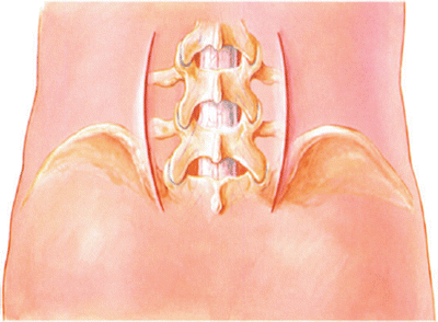
The most commonly used approaches to this area are the direct paraspinous muscle-splitting incision and the more lateral approach, which elevates and dissects under the lumbosacral muscle (Fig. 49.1). The muscle-splitting incision affords not only alar transverse exposure for fusions but also excellent exposure for laminectomy1,2. The more lateral approach removes a bicortical portion of the posterior superior iliac spine, attempts to preserve muscle and fascial integrity, and allows excellent exposure of the tips of the transverse processes to the facet joints.3,4 There is probably no great advantage to preserving the denervated muscle with the more lateral approach. We recommend the more lateral approach for alar transverse fusion and the muscle-splitting incision when laminectomy and root exploration are needed in addition to fusion.


Fig. 49.1
The skin incisions on the bilateral paraspinous approach to the lower lumbar spine may be one of two types: (1) a lateral incision three fingerbreadths from the midline designed to include the iliac crest in the wound while the iliac crest is removed and dissection performed under the paraspinous musculature from lateral to medial; (2) a more medial paraspinous approach designed for muscle splitting and direct approach to the facet joint and transverse processes without including the iliac crest through that incision. A midline incision may be used for a bilateral paraspinous fusion. The incision must be long to allow adequate lateral retraction to help develop the incision in the lumbodorsal fascia and lateral approach to the paraspinous musculature
1.
Position the patient prone on the table with an appropriate operating frame.
2.
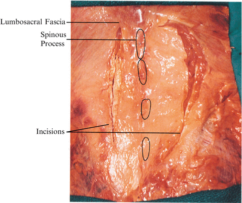
After adequate prepping and draping, make a curved paraspinous incision approximately three fingerbreadths from the midline, L4 to S2 (Fig. 49.2).


Fig. 49.2
The paravertebral fascia incisions
3.
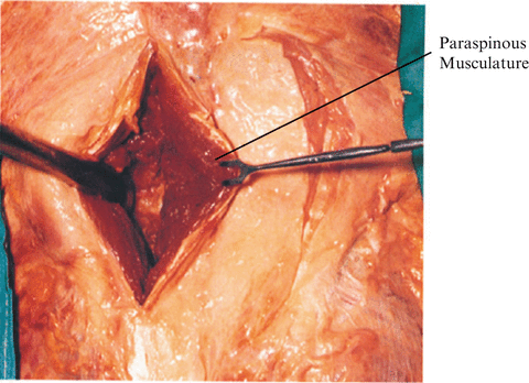
Cut through skin and subcutaneous tissue to the lumbar fascia. The posterior superior iliac spine is in the inferior portion of the wound. Palpate the transverse process of L5 and L4. Remove the fascial insertions on the iliac crest and reflect medially with a small sliver of bone (Fig. 49.3).


Fig. 49.3
The more medial paraspinous muscle-splitting incision. Open the fascia and dissect with your finger directly through the muscle to identify the L5–S1 facet, the ala of the sacrum lateral to that facet, and the L4–L5 joint cephalad. Remove the muscle attachments to the facets and transverse processes from L4 to the sacrum
4.
With an elevator, dissect subperiosteally both the inner and outer tables of the posterior iliac crest to a depth of 2 to 3 cm.
5.
Osteotomize the ilium with an osteotome, beginning at the medial portion of the posterosuperior iliac spine not involving the sacral iliac joint and extending approximately 5 to 6 cm on the iliac crest.
Caution: The clunial nerves will be in the lateral aspect of the wound and must be avoided. A safe margin of error is approximately one handbreadth lateral to the posterosuperior iliac spine 1.
6.
Get Clinical Tree app for offline access

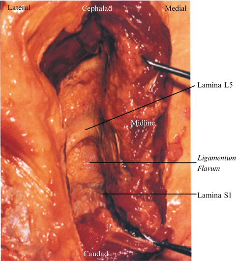
Remove the bicorticate iliac spine graft. The incision in the paraspinous fascia is extended cephalad on the lateral border facet to the proximal level of the L4 transverse process. Medially, the interlaminar areas can be exposed for a canal decompression or a laminectomy (Fig. 49.4).


Fig. 49.4


If a decompression is to be done, dissect more medially and clear the laminae of soft tissue
Stay updated, free articles. Join our Telegram channel

Full access? Get Clinical Tree


