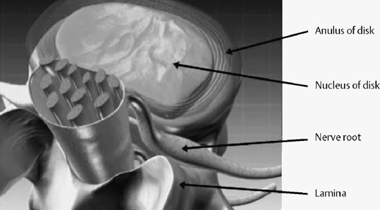
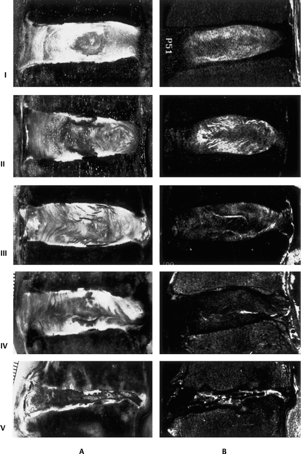
III 10 Biochemical Aspects of Intervertebral Disk Degeneration 11 Degenerative Cervical Spine Disorders: Surgical and Nonsurgical Treatment 12 Degenerative Thoracic Spine Conditions 13 Lumbar Disk Disease: Pathogenesis and Treatment Options 15 Surgical Management of Axial Back Pain I. Intervertebral disk A. Cells 1. Notochordal cells present embryologically disappear by adult life 2. Chondrocyte-like cells in the nucleus pulposus and annulus fibrosus most likely originate from chondrocytes in the cartilaginous end plate. 3. No significant cell turnover 4. Apoptosis with aging and disk degeneration B. Gross structures (from peripheral to central) (Fig. 10–1) 1. Outer fibrous annulus fibrosus a. Primarily collagen fibrils in oblique layers b. Limited vascular and nerve supply c. Sinuvertebral nerve posteriorly d. Sympathetic fibers anteriorly 2. Inner annulus fibrosus a. Fibrocartilaginous 3. Transition zone a. Thin zone of fibrous tissue between inner annulus and the nucleus pulposus 4. Nucleus pulposus C. Matrices 1. Collagens a. Annulus (70%) (1) Predominantly type I (2) Type I, II, III, V, VI, IX, XI b. Nucleus (20%) (1) Predominantly type II (2) Type II, VI, IX, and XI c. Provide tensile strength d. Collagen cross-linking by covalent bonds via modification of lysine/hydroxylysine residues e. Highest concentration of cross-linking in nucleus D. Disk degeneration 1. Collagen synthesis and content increase in the nucleus 2. Decreased concentration of cross-linking in the annulus E. Proteoglycans (PGs) 1. PG aggregates’ constituents a. Central hyaluronan filament b. Link proteins attach multiple glycosoaminoglycan molecules c. Large PGs (1) Aggrecan (a) Similar to articular cartilage (b) Half the size of PGs found in cartilage (c) Higher keratin sulfate:chondroitin sulfate ratio (d) Higher molecular weights of keratin sulfate (e) Increased hyaluron content (f) Important in water retention (g) Provides compressive strength d. Small PGs (1) Biglycan, decorin, lumican, fibromodulin (2) Involved in organization of collagen and fibril formation (3) PG content and synthesis vary depending on age, region, and degeneration. (4) PG synthetic activity of adult normal annulus approximately one third lower than articular cartilage and young nucleus (5) Synthetic activity greatest in the inner annulus F. Aging and degeneration (Fig. 10–2) 1. Keratin sulfate:chondroitin sulfate ratio increases
Degenerative Spinal Conditions
10
Biochemical Aspects of Intervertebral Disk Degeneration

Neupsy Key
Fastest Neupsy Insight Engine


