Fig. 7.1
The three-dimensional finite element method (3D-FEM) model of the spinal cord consisted of gray matter, white matter, and pia mater
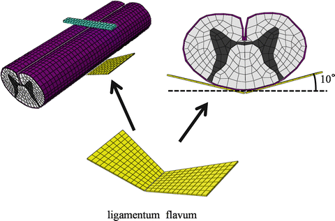
Fig. 7.2
The ligamentum flavum model was established using the rear of the spinal cord

Fig. 7.3
The shapes for anterior compression of the spinal cord were central type, lateral type, and diffuse type
The model simulated CSM under three shapes of anterior compression of the spinal cord, as well as dynamic compression under neck extension of the cervical spinal cord. The ligamentum flavum was located at the anterior surface of the cephalad lamina and at the superior margin of the caudal lamina.
The spinal cord consists of three distinctive materials referred to as white matter, gray matter, and pia mater. The mechanical properties (Young’s modulus and Poisson’s ratio) of the gray and white matter were determined using data obtained by the tensile stress strain curve and stress relaxation under various strain rates [10, 11]. The mechanical properties of pia mater and the ligamentum flavum were obtained from the literature [12, 13].
For the simulation of CSM under neck extension, the extension angle of the superior margin of spinal cord was 20°. Based on the assumption that the caudal vertebral body of the responsible lesion in CSM patients was stable, the bottom of the ligamentum flavum was constrained in all directions. The superior margin of the ligamentum flavum shifted distally according to the movement of cephalad lamina. The angle of neck extension and the direction in which the ligamentum flavum moved were obtained from each angle of C3–C4 kinematic MRI published by Iwabuchi et al. [14] (Fig. 7.4).
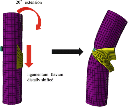

Fig. 7.4
For the simulation of neck extension, an extension of 20° was applied to the spinal cord. The ligamentum flavum moved distally in accordance with movement of the cephalad lamina
FEM analysis was conducted under each of the above conditions and in duplicate to test the reproducibility of results with this model.
7.2.2 Stress Distribution
When the shape of the spinal cord resulting from anterior compression was the central type, stress distributions were observed in the posterior horn and in parts of the posterior funiculus, lateral funiculus, and anterior horn beneath the invagination by ligamentum flavum. Under anteroposterior compression, high stress distributions were observed in the gray matter, anterior funiculus, and part of the lateral funiculus (Fig. 7.5a).
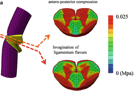
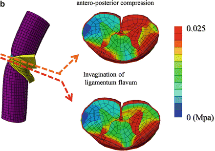
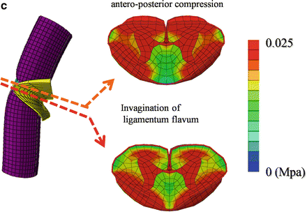


Fig. 7.5
Stress distributions under invagination of the ligamentum flavum and under anteroposterior compression are shown for the central-, lateral-, and diffuse-type shapes following anterior compression of the spinal cord (a–c)
When the shape of the spinal cord resulting from anterior compression was the lateral type, stress distributions were observed in the gray matter and lateral funiculus on the compression side, as well as parts of the posterior funiculus and posterior horn on the non-compression side beneath the invagination by ligamentum flavum. Under anteroposterior compression, high stress distributions were observed in the gray matter, anterior funiculus, and lateral funiculus on the compression side (Fig. 7.5b).
When the shape of the spinal cord resulting from anterior compression was the diffuse type, stress distributions were observed in the gray matter, lateral funiculus, and posterior funiculus beneath the invagination by ligamentum flavum. Under anteroposterior compression, high stress distributions were observed in the gray matter, anterior funiculus, lateral funiculus, and part of the posterior funiculus (Fig. 7.5c).
7.3 Discussion
Although it is the most common reason for spinal cord dysfunction in the elderly, CSM has no pathognomonic symptoms or physical signs. Because no single neurologic feature is unique to cervical myelopathy, diagnosis must be established by the affirmation of associated clinical signs and symptoms, in parallel with the exclusion of other clinical entities that may also show these signs.
CSM is a well-described clinical syndrome that can arise due to a combination of etiological factors. This clinical entity manifests itself following static or dynamic compression of the spinal cord, causing compression of the enclosed cord and leading to local tissue ischemia, injury, and neurologic impairment. Static factors include developmental canal stenosis, bulging of the intervertebral disc posterior margin, and hypertrophy of the ligamentum flavum. With regard to dynamic factors, the spinal cord of CSM patients is likely to be compressed during neck extension because of the protrusion of ligamentum flavum and intervertebral discs into the spinal canal (pincer mechanism) [2]. Recent developments in imaging have enabled closer examination of the cervical spine through improved CT and MRI. Using multidetector-row CT, Machino et al. reported narrowing of the spinal cord cross-sectional area during neck extension in patients with CSM [7]. In healthy individuals, Muhle et al. found that the subarachnoid space was smallest at extension compared to its size at mid-position and at flexion of the cervical spine. These workers also showed an increasing prevalence of functional cord impingement from the posterior aspect and from both the anterior and posterior aspects during neck extension in patients with advancing stages of degenerative disease of the cervical spine [15, 16].
Stay updated, free articles. Join our Telegram channel

Full access? Get Clinical Tree


