Figure 8.1
(a) This image depicts a gross, intra-operative view of a cerebral aneurysm in the center of the picture. At the sides of the aneurysms (red color) is the brain tissue. (b) This image shows a metallic clip about to be placed around the aneurysm (surgical clipping), sealing it off from the circulation
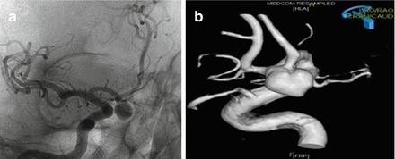
Figure 8.2
(a) 65 year-old with a large anterior communicating artery aneurysm with a cerebral angiogram of cerebral arteries when viewed from the front, showing a large aneurysm in the anterior communicating portion of the anterior cerebral arteries. (b) 3D reconstruction software takes pictures from a cerebral angiogram and creates an image similar to this, to better characterize the size and location of the aneurysm
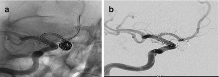
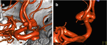
Figure 8.4
(a) 61 year-old female with subarachnoid hemorrhage secondary to ruptured anterior communicating aneurysm. 3D reconstruction of large anterior communicating artery aneurysm when viewed from the right side. (b) 3D reconstruction of large anterior communicating artery aneurysm when viewed from the left side
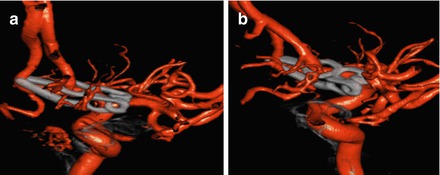
Figure 8.5
(a) The aneurysm in Fig. 8.4 has now been treated with a clip (front view). Note the jaws of the clip have an “open” portion that allows it to wrap around one of the anterior cerebral arteries without occluding it and still seal off the aneurysm (***keep image aspect ratio when enlarge***). (b) Same aneurysm after clip ligation when viewed from the side
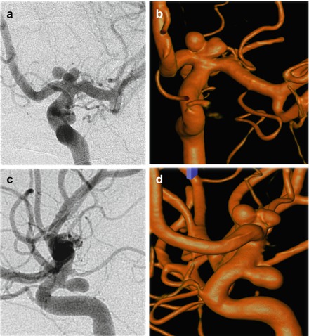
Figure 8.6
(a) 28 year-old male with subarachnoid hemorrhage found to have multiple left internal carotid artery aneurysms. Cerebral angiogram (front view) showing bilobed aneurysm at the very end of the internal carotid artery where it bifurcates into the anterior and middle cerebral arteries as well as a more proximal aneurysm projecting backwards off the internal carotid artery. (b) 3D reconstruction of the cerebral angiogram again showing the bilobed aneurysm at the end of the internal carotid artery. The aneurysm projecting backwards is not easily seen in this view. (c) Cerebral angiogram (side view) showing more clearly the aneurysm that projects backwards off the internal carotid artery. (d) 3D reconstruction of the cerebral angiogram (side view) showing both the bilobed aneurysm at the end of the internal carotid artery and the more proximal aneurysm that projects backwards off the internal carotid artery
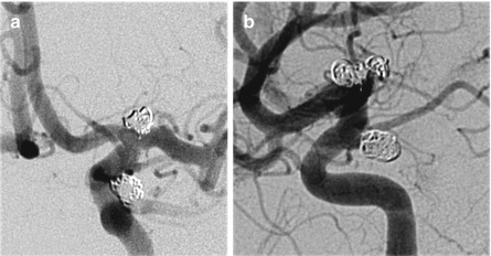
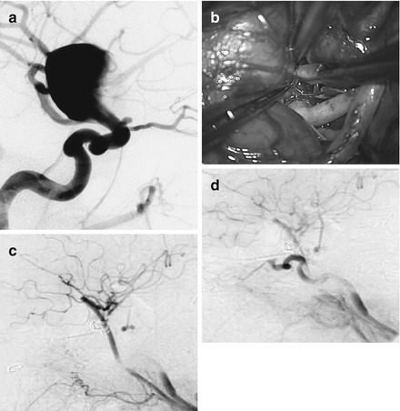
Figure 8.8


(a) 63 year-old woman suffered ruptured giant carotid aneurysm, angiogram in lateral view. This aneurysm cannot be treated with either coiling or direct clipping. (b) Intra-operative photo of bypass (pink tubular structure in right corner) to provide alternate blood flow into the brain from neck; followed with permanent aneurysm occlusion. (c) Intra-operative angiogram to show the bypass (dark lower center vessel) supplying the whole left brain. (d) Intra-operative angiogram to show complete occlusion of the aneurysm
Related posts:


Stay updated, free articles. Join our Telegram channel
Full access? Get Clinical Tree
