Brainstem Anatomy
A. See also
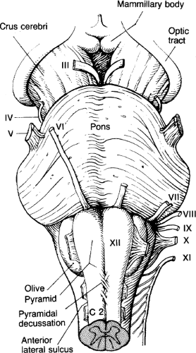 Figure 3. The ventral surface of the brainstem. (From Duus P. Topical Diagnosis in Neurology. New York: Thieme, 1983:101, with permission.) |
B. Slice anatomy
For easier comparison with MRIs, the five images in Figure 4 are in radiologic convention, as if looking up from the feet, upside down and backwards from neuropathologic convention. In order, they show the upper medulla, lower pons, midpons, upper pons, and midbrain.
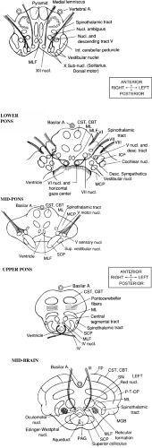 Figure 4. Brainstem cross-sections drawn in radiologic convention. (From Schwamm L. In Batjer HH, ed. Cerebrovascular Disease. Philadelphia: Lippincott, 1996, with permission.) Continued on next page. |
Related posts:


Stay updated, free articles. Join our Telegram channel

Full access? Get Clinical Tree


