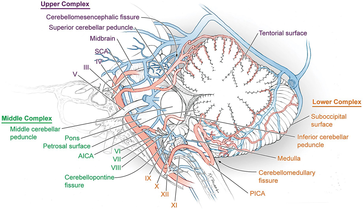
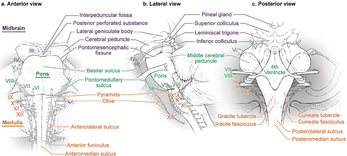
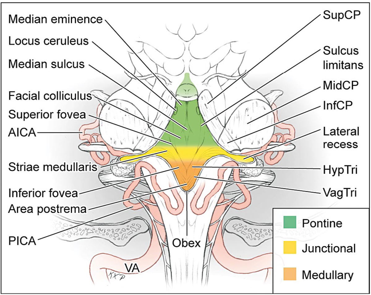
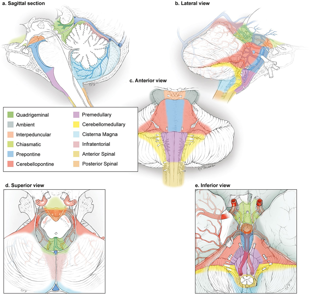
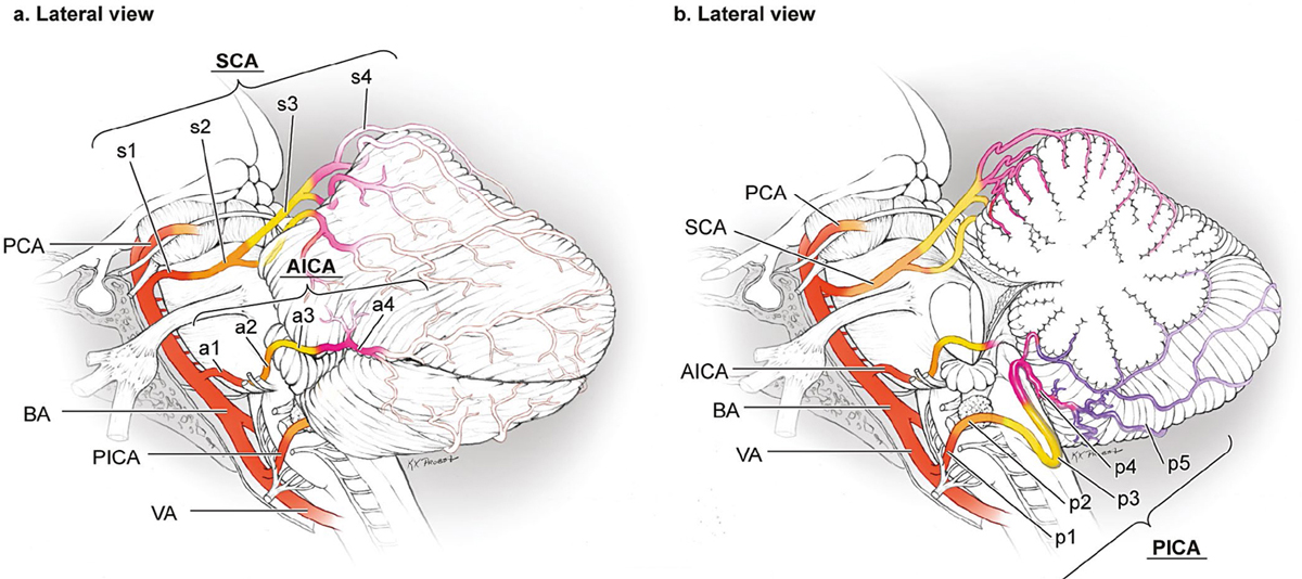
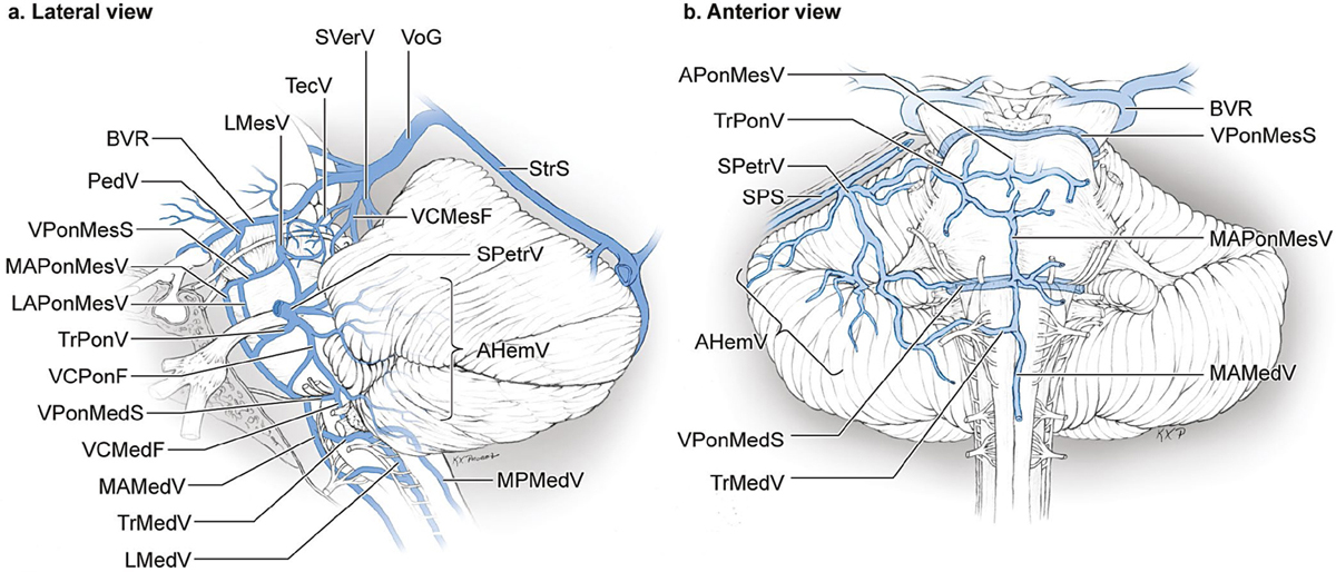
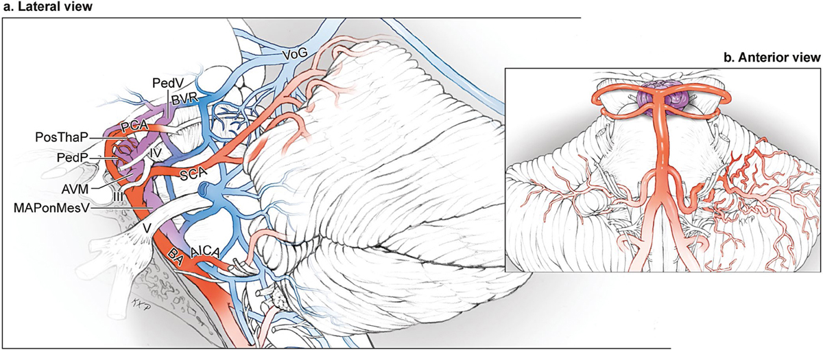
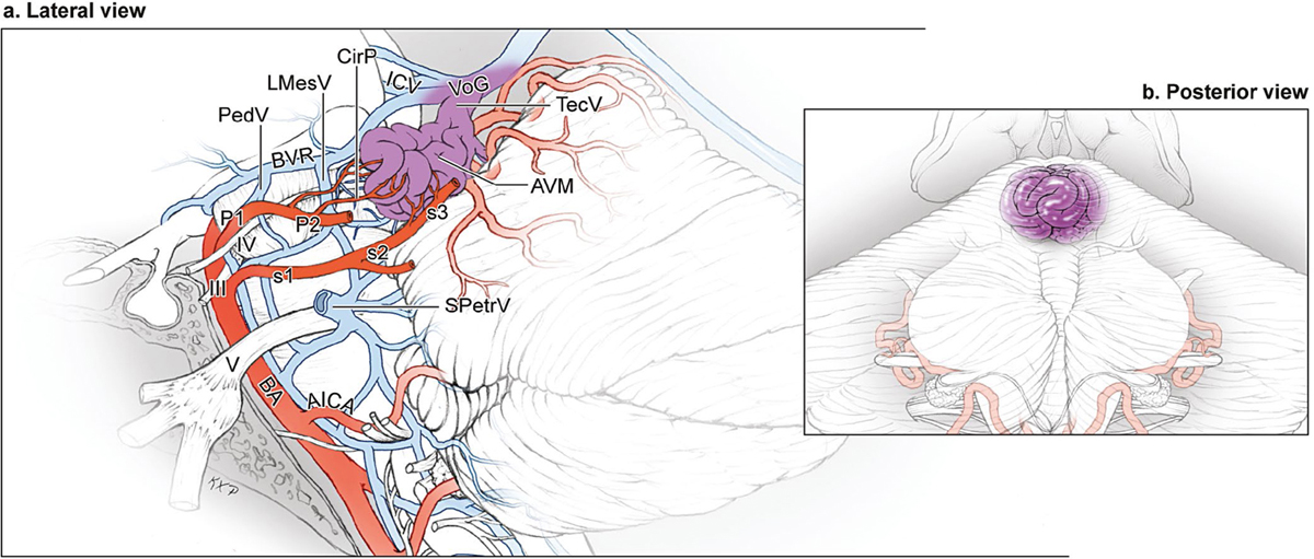
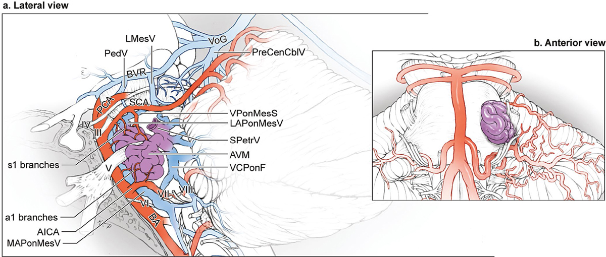
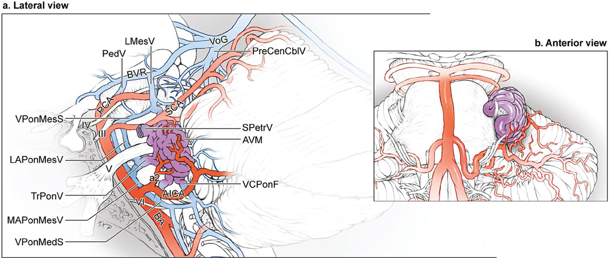
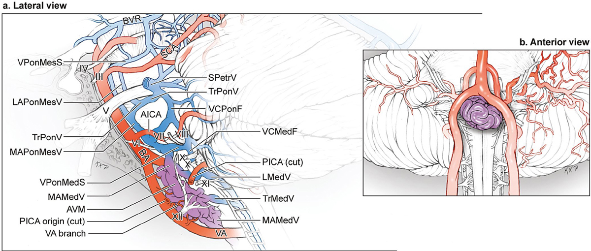
16 Brainstem Arteriovenous Malformations Structures in the posterior fossa occur in “3’s.” The brainstem is composed of midbrain, pons, and medulla; superior, middle, and inferior cerebellar peduncles connect the brainstem and cerebellum; brainstem and cerebellum are divided by cerebellomesencephalic, cerebellopontine, and cerebellomedullary fissures; the cerebellum has tentorial, petrosal, and suboccipital surfaces; and brainstem and cerebellum are supplied by the superior cerebellar artery, anterior inferior cerebellar artery, and posterior inferior cerebellar artery. Three neurovascular complexes are defined with one of the three brainstem parts, one of the three cerebellar surfaces, one of the three peduncles, one of the three major fissures, and one of the three cerebellar arteries (Fig. 16.1). In addition, each neurovascular complex contains a group of cranial nerves: the upper complex contains the oculomotor, trochlear, and trigeminal nerves; the middle complex contains the abducens, facial, and vestibulocochlear nerves; and the lower complex contains the glossopharyngeal, vagus, accessory, and hypoglossal nerves. In summary, the upper complex includes the superior cerebellar artery (SCA), midbrain, cerebellomesencephalic fissure, superior cerebellar peduncle, tentorial cerebellar surface, and cranial nerves (CN) 3/4/5. The SCA arises in front of the midbrain and passes below CN3 and CN4 and above CN5 to reach the cerebellomesencephalic fissure, where it runs on the superior cerebellar peduncle and terminates by supplying the tentorial surface. The middle complex includes the anterior inferior cerebellar artery (AICA), pons, middle cerebellar peduncle, cerebellopontine fissure, petrosal surface, and CN6/7/8. The AICA arises at the pontine level, courses under CN6 and along CN7/8 to reach the surface of the middle cerebellar peduncle, where it courses along the cerebellopontine fissure and terminates by supplying the petrosal surface. The lower complex includes the posterior inferior cerebellar artery (PICA), medulla, inferior cerebellar peduncle, cerebellomedullary fissure, suboccipital surface, and CN9/10/11/12. The PICA arises at the medullary level, encircles the medulla, passing in relationship to lower CNs to reach the surface of the inferior cerebellar peduncle, where it dips into the cerebellomedullary fissure and terminates by supplying the suboccipital surface. Fig. 16.1 Microsurgical anatomy of the three neurovascular complexes of the posterior fossa [upper (purple), middle (green), and lower (orange) complexes], each defined by a brainstem part, cerebellar surface, cerebellar peduncle, major fissure, cerebellar artery, and cranial nerve bundle (lateral view with sagittal cross section through the vermis). Surface anatomy provides landmarks for brainstem arteriovenous malformations (AVMs) (Fig. 16.2). Midbrain landmarks include the cerebral peduncles, CN3, interpeduncular fossa, posterior perforated substance, and pontomesencephalic fissure on the anterior surface; the cerebral peduncle, lateral geniculate body (LGB), lemniscal trigone, and CN4 on the lateral surface; and the pineal gland, tectum (superior and inferior colliculi), and CN4 on the posterior surface. Pontine landmarks include the basilar sulcus, pontomedullary sulcus, and CN6 on the anterior surface; the middle cerebellar peduncle, CN5 (sensory and motor roots), CN7/8 on the lateral surface; and the upper half of the fourth ventricular floor (above stria medullaris) on the posterior surface. Medullary landmarks include the pyramids, anteromedian sulcus between the pyramids, anterolateral sulcus, and CN12 rootlets arising from the anterolateral sulcus on the anterior surface; the olive, anterolateral (preolivary) sulcus, posterolateral (postolivary) sulcus, and CN9/10/11 rootlets arising from posterolateral sulcus on the lateral surface; and the posteromedian sulcus, gracile fasciculus and tubercle medially, cuneate fasciculus and tubercle laterally, posterior intermediate sulcus between them, inferior cerebellar peduncles, and the lower half of the fourth ventricular floor (below stria medullaris) on the posterior surface. Fig. 16.2 Surface anatomy of the midbrain (purple), pons (green), and medulla (orange), seen in (a) anterior, (b) lateral, and (c) posterior views. The fourth ventricular floor (a.k.a., rhomboid fossa) has three parts (Fig. 16.3): a pontine part extending from the cerebral aqueduct triangularly down to an imaginary line connecting the lower margin of the cerebellar peduncles; a junctional part forming a strip between the lateral recesses; and a medullary part extending triangularly down to the obex. Surface landmarks in the fourth ventricular floor include the median sulcus, facial colliculus, superior fovea, locus ceruleus, stria medullaris, inferior fovea, hypoglossal triangle, vagal triangle, and area postrema. The median sulcus divides the floor longitudinally in the midline, and the median eminence is a raised strip that parallels the median sulcus. The median eminence contains the facial colliculus rostrally and three triangular areas caudally—the hypoglossal triangle, vagal triangle, and area postrema—collectively forming the calamus scriptorius, named for its resemblance to a feather or pen nib. The sulcus limitans is another longitudinal sulcus on the lateral border of the median eminence. The locus ceruleus is a bluish gray area at the rostral end of the sulcus limitans in the lateral margin of the floor, and the superior and inferior foveae are dimples in the sulcus limitans, the superior one just lateral to the facial colliculus in the pons and the inferior one just lateral to the hypoglossal trigone in the medulla. The facial colliculus overlies the abducens nucleus and the ascending root of CN7. The hypoglossal trigone overlies the hypoglossal nucleus, and just below it, the vagal trigone overlies the dorsal nucleus of the vagus nerve. The area postrema is at the lower limit of the median eminence immediately rostral to the obex. The striae medullaris, which are white strands traversing the junctional part of the floor from the lateral recess to the sulcus limitans, overlie the vestibular area and the underlying vestibular nuclei. Fig. 16.3 Surface anatomy of the fourth ventricular floor, which forms the posterior surface of the pons and half of the medulla. Liliequist’s membrane separates the chiasmatic and interpeduncular cisterns and demarcates the boundary between the supra- and infratentorial cisterns (Fig. 16.4). The mid-brain is surrounded by the interpeduncular, ambient, and quadrigeminal cisterns. The pons is surrounded by the prepontine and cerebellopontine cisterns. The medulla is surrounded by the premedullary and cerebellomedullary cisterns, and the cisterna magna. Midline cisterns are unpaired (IpC, PonC, MedC, QuadC, and MagnC), whereas lateral cisterns are paired (AmbC, CbPonC, and CbMedC). Anterior and posterior spinal cisterns surround the cord below the foramen magnum. Fig. 16.4 Microsurgical anatomy of the posterior fossa cisterns: (a) sagittal cross-sectional, (b) lateral, (c) anterior, (d) superior, and (e) inferior views. The midbrain is surrounded by the interpeduncular, ambient, and quadrigeminal cisterns. The pons is surrounded by the prepontine and cerebellopontine cisterns. The medulla is surrounded by the premedullary and cerebellomedullary cisterns, and the cisterna magna. The SCA is divided into four segments (Fig. 16.5): anterior pontomesencephalic, lateral pontomesencephalic, cerebellomesencephalic, and cortical segments. The anterior pontomesencephalic segment (s1) begins at the SCA origin, courses underneath CN3, and ends at the anterolateral brainstem, medial to the tentorial edge. The lateral pontomesencephalic segment (s2) begins at the anterolateral brainstem, dips caudally to the trigeminal root, and terminates at the entrance to the cerebellomesencephalic fissure. The cerebellomesencephalic segment (s3) courses posteriorly within this fissure, following CN4 and the superior cerebellar peduncle. A series of hairpin curves leads deep into the fissure and then upward to reach the anterior edge of the tentorium. The cortical segment (s4) begins as distal branches exit the cerebellomesencephalic fissure and supply the cerebellum’s tentorial surface. The AICA is divided into four segments (Fig. 16.5): anterior pontine, lateral pontine, flocculopeduncular, and cortical segments. The anterior pontine segment (a1) begins at the AICA’s origin from the basilar trunk and ends at the level of a line drawn through the long axis of the inferior olive extended upward on the pons. This segment courses below CN6 as it ascends to Dorello’s canal. The lateral pontine segment (a2) begins at the anterolateral margin of the pons and passes through the cerebellopontine angle alongside CN7/8, the internal auditory meatus, lateral recess, and choroid plexus protruding from the foramen of Luschka. The labyrinthine artery, recurrent perforating arteries, and subarcuate artery arise from this segment. The flocculopeduncular segment (a3) begins as the artery passes the flocculus and middle cerebellar peduncle, and ends as it reaches the cerebellopontine fissure. It commonly bifurcates near CN7/8 into rostral and caudal trunks. The cortical segment (a4) supplies the petrosal surface distal to the cerebellopontine fissure. The PICA has five segments (Fig. 16.5): anterior medullary, lateral medullary, tonsillomedullary, telovelotonsillar, and cortical segments. The anterior medullary segment (p1) begins at the PICA’s origin, lies anterior to the medulla, and extends past the hypoglossal rootlets at the medial edge of the inferior olive. The lateral medullary segment (p2) extends from the most prominent point of the inferior olive to the rootlets of CN9/10/11 at the lateral edge of the olive. The tonsillomedullary segment (p3) begins where the PICA passes under or between the rootlets of CN9/10/11, descends to the inferior pole of the cerebellar tonsil, reverses course in the caudal or infratonsillar loop, and ascends along the medial tonsil to its midpoint. The telovelotonsillar segment (p4) begins at the midpoint of the PICA’s ascent, continues toward the roof of the fourth ventricle, turns around again to form a cranial or supratonsillar loop, and courses downward and posteriorly to the tonsillobiventral fissure. This segment derives its name from its branches to the choroid plexus of the fourth ventricle (tela choroidea) and its association with the roof of the fourth ventricle (inferior medullary velum). Cortical segment (p5) begins as the PICA emerges from the tonsillobiventral fissure where the tonsil, vermis, and biventral lobule meet. This segment has a medial trunk supplying the vermian surface and a lateral trunk supplying the tonsillar and hemispheric surfaces. Fig. 16.5 Microsurgical anatomy of the brainstem and cerebellar arteries. Brainstem surfaces are supplied by the first three segments of the cerebellar arteries, with the anterior surfaces supplied by the first segments (s1 anterior pontomesencephalic, a1 anterior pontine, and p1 anterior medullary segments), the lateral surfaces supplied by the second segments (s2 lateral pontomesencephalic, a2 lateral pontine, and p2 lateral medullary segments), and the posterior surfaces supplied by the third segments (s3 cerebellomesencephalic, a3 flocculopeduncular, and p3 tonsillomedullary segments), shown in lateral views (a) with and (b) without the left cerebellum (sagittal section through the vermis). Brainstem veins are named according to the division of the brainstem, the surface, and their course longitudinally or transversely (Fig. 16.6). Longitudinal veins include the median anterior pontomesencephalic vein (MAPonMesV), median anterior medullary vein (MAMedV), lateral anterior pontomesencephalic vein (LAPonMesV), lateral anterior medullary vein (LAMedV), lateral mesencephalic vein (LMesV), lateral medullary vein (LMedV), and retro-olivary veins (ReOlvV). Transverse veins are the vein of the pontomesencephalic sulcus (VPonMesS), vein of the pontomedullary sulcus (VPonMedS), transverse pontine vein, transverse medullary vein, peduncular vein, and posterior communicating vein (PComV). Veins draining the brainstem bridge across the subarachnoid spaces to reach the dural sinuses. The peduncular, PComV, and tectal vein (TecV), as well as the longitudinal veins, ascend to the basal vein of Rosenthal (BVR) en route to the vein of Galen (VoG). Anterior brainstem veins course laterally to the superior petrosal (SPetrV, or Dandy’s vein) and inferior petrosal veins (IPetrV) that drain to the petrosal sinuses (SPS and IPS, respectively). The brainstem is one of the inviolable areas packed densely with cranial nerve nuclei and tracts. All brainstem AVMs are eloquent and drain deeply, raising their Spetzler-Martin grades to at least grade III. Fortunately, brainstem AVMs are rare (4% of AVMs in my surgical experience [25 patients] and 2% to 6% in published series) and many sit on the brainstem surface rather than in the parenchyma (70% in my surgical experience). This pial or “epipial” location makes them different from cerebral and cerebellar AVMs. The categorization of brainstem AVMs is based on the brainstem level and surface, as with other AVMs. Half were located in pons, one fourth in the midbrain, and one fourth in the medulla. Fig. 16.6 Microsurgical anatomy of the brainstem veins. (a) Veins of the lateral brainstem (lateral view). (b) Veins of the anterior brainstem and cerebellum (anterior view). Brainstem veins are named according to the division of the brainstem (mesencephalon, pons, or medulla), the surface (anterior, lateral, or posterior), and their course (longitudinal or transverse). Fig. 16.7 The anterior midbrain AVM: (a) lateral and (b) anterior views. This AVM is located on the anterior midbrain surface on, in, or between the cerebral peduncles, is supplied by P1 PCA branches (PosThaP and PedP), and is drained by anterior veins (MAPonMesV, PedV, and PComV). The anterior midbrain AVM is located on the anterior mid-brain surface on, in, or between the cerebral peduncles (Fig. 16.7). This subtype is associated with CN3 and lies behind the P1 posterior cerebral artery (PCA) and PosThaP in the interpeduncular fossa. Portions of the AVM sit in the interpeduncular cistern. Perforating arteries from both the P1 PCA supply the nidus (PosThaP and PedP), and it is drained by MAPonMesV, PedV, and PComV, which then collect in the BVR. The posterior midbrain AVM sits on the tectum/quadrigeminal plate, below the pineal gland and above the superior cerebellar peduncles, often intimate with the trochlear nerves (Fig. 16.8). Portions of the AVM reside in the quadrigeminal cistern. These AVMs are fed by the long circumflex perforator (CirP) from the P1 and P2 PCA segments, as well as branches from the s3 SCA, typically bilaterally. Venous drainage into the tectal vein and the vein of the cerebellomesencephalic fissure (VCMesF) collect in the VoG. Although the lateral surface of the midbrain is sizable, no lateral midbrain AVM was encountered. Fig. 16.8 The posterior midbrain AVM: (a) lateral and (b) posterior views. This AVM is located on the posterior midbrain surface on or adjacent to the quadrigeminal plate. It is supplied bilaterally by the PCA (long circumflex perforators from P1 and P2 segments) and the SCA (s3 branches), and is drained by veins ascending to the vein of Galen (TecV and VCMesF). The anterior pontine AVM sits on the rectangular surface between the basilar sulcus medially and the CN5 root laterally, and between the pontomesencephalic sulcus superiorly and the pontomedullary sulcus inferiorly (Fig. 16.9). These exophytic AVMs protrude into the prepontine and cerebellopontine cisterns. Despite its anterior location, this AVM is unilateral and does not cross the midline. It is located between the s1 SCA superiorly and the a1 AICA inferiorly, and is supplied by both arteries. Enlarged perforating branches from the basilar trunk also feed this AVM directly. Interestingly, arteries from the meningohypophyseal trunk of the internal carotid artery (ICA) travel through Meckel’s cave along CN5 and can contribute to these AVMs (three cases). Venous drainage collects medially in MAPonMesV or more commonly in SPetrV and SPS laterally. These AVMs are intimately associated with CN5, often entangled in nerve fibers. The lateral pontine AVM sits on the triangular surface between the CN5 root medially and the V-shaped cerebellopontine fissure laterally, as the pons transitions into the middle cerebellar peduncle (Fig. 16.10). The extent of pial invasion varies among these AVMs, with some lying on the pial surface and others penetrating deeply into the middle cerebellar peduncle. Surprisingly, this portion of the brachium pontis tolerates surgical transgression, making intraparenchymal lateral pontine AVMs resectable lesions. They are supplied by the AICA (a2 and a3 segments) and, unlike anterior pontine AVMs, do not receive input from the SCA. Venous drainage collects in SPetrV and SPS. Lateral pontine AVMs are located in the cerebellopontine angle and more medial than petrosal cerebellar AVMs, which are also in the cerebellopontine angle but based on the petrosal surface in the cerebellar lobules rather than in the middle cerebellar peduncle. Lateral pontine AVMs were the most common brainstem subtype (27%). No posterior pontine AVM was encountered. Fig. 16.9 The anterior pontine AVM: (a) lateral and (b) anterior views. This AVM is located on the rectangular surface between the basilar sulcus medially, the trigeminal root laterally, and the pontomesencephalic and pontomedullary sulci. It is supplied unilaterally by s1 SCA and a1 AICA branches, as well as direct branches from the basilar trunk, and drains to MAPonMesV medially or SPetrV laterally. Fig. 16.10 The lateral pontine AVM: (a) lateral and (b) anterior views. This AVM is located on the triangular surface of the pons and brachium pontis between the CN5 root medially and the V-shaped cerebellopontine fissure laterally. It is supplied unilaterally by the a2 and a3 AICA branches and is drained by SPetrV. The anterior medullary AVM is located on the anterior surface below the pontomedullary sulcus, between the anterolateral sulci and anterior to the hypoglossal nerve rootlets (Fig. 16.11). Portions of the AVM reside in the premedullary cistern. This AVM sits posteroinferior to the vertebrobasilar junction (VBJ) in the midline and is supplied bilaterally by vertebral artery (VA) branches at the level of the anterior spinal artery origins (distal V4 segment). The PICA originates lateral to this area and does not contribute. Venous drainage is through MAMedV. The lateral medullary AVM is located below the pontomedullary sulcus, lateral to the anterolateral (preolivary) sulcus and posterior to the hypoglossal nerve rootlets (Fig. 16.12). This AVM is visible on the lateral medullary surface through a far lateral craniotomy in the location of the PICA’s p2 segment. It tends to be small in size and respects the pia, with most of the AVM in the cerebellomedullary cistern. This AVM is unilateral and fed by small branches directly from the VA and PICA. It is drained by LMedV and MAMedV. Lateral medullary AVMs were more common than the anterior medullary AVM (19% vs. 4%, respectively). No posterior medullary AVMs were encountered. Fig. 16.11 The anterior medullary AVM: (a) lateral and (b) anterior views. This AVM is located below the pontomedullary sulcus, between the anterolateral sulci, and anterior to hypoglossal nerve rootlets. It is supplied by branches from the distal vertebral artery proximal to or at the vertebrobasilar junction and is drained in the midline by MAMedV and MAPonMesV.
 Microsurgical Anatomy
Brain
Arteries
Veins
 Six Brainstem AVM Subtypes
The Anterior Midbrain AVM
The Posterior Midbrain AVM
The Anterior Pontine AVM
The Lateral Pontine AVM
The Anterior Medullary AVM
The Lateral Medullary AVM
Stay updated, free articles. Join our Telegram channel

Full access? Get Clinical Tree


