Fig. 6.1
Non-granulomatous encephalitis. A hematoxylin and eosin (H&E)-stained brain section shows scattered lymphocytic infiltrates and perivascular lymphocytic cuffing (From Erdem et al. [13], with permission)
6.4 Diagnosis
The diagnosis requires the combination of several approaches. All patients must be questioned about epidemiological risk factors for brucellosis. Clinical examination, routine hematological and biochemical laboratory tests, and radiological investigations are elements of the primary approach.
The diagnosis of brucellar meningoencephalitis requires the demonstration of the findings related to meningeal inflammation, abnormal CSF findings, and direct or indirect evidence of Brucella in the CSF [20, 25, 29].
Lumbar puncture is suggested in all cases with symptoms and clinical findings consistent with neurobrucellosis, unless contraindicated. Analysis findings of CSF reveal lymphocytic pleocytosis, increased CSF protein level, and normal or decreased CSF/plasma glucose ratio [31].
Culture, when positive, is accepted as the gold standard for the laboratory diagnosis of brucellosis. Importantly, the speed of recovery is dependent on the culture method used and the type of specimen used. In this regard, the conventional method requiring a long incubation time of 6 weeks has a yield of 5–20 % in chronic, focal, and complicated cases [2]. In a study automated culture systems yielded the pathogen from the CSF significantly higher than the conventional culture [11].
As culturing of Brucella takes time, laboratory diagnosis very often relies on detecting specific serum antibodies. In the serological diagnosis of brucellosis, Rose Bengal test (RBT), microagglutination test, STA, indirect Coombs (antihuman globulin) test, complement fixation, enzyme-linked immunosorbent assay (ELISA), and immunocapture agglutination techniques have been used [2, 15].
Serological tests such as serum STA, RBT, and ELISA as well as CSF RBT, STA, and ELISA were applied to 177 patients with chronic brucellar meningitis or meningoencephalitis in a multicenter, retrospective study [11]. The sensitivities of the tests were 94 % for serum STA, 96 % for serum RBT, 78 % for CSF STA, and 71 % for CSF RBT [11]. No significant difference was detected between the sensitivities of CSF RBT and CSF STA (p = 0.163) and the serum RBT and the serum STA (p = 0.500), whereas the differences for either serum and CSF RBT or serum and CSF STA were significant statistically (p < 0.001) [11].
A total of 31 neurobrucellosis cases, 14 with meningoencephalitis, were evaluated in a study. STA was performed on 16 CSF samples; 14 samples were found positive ranging from 1/20 to 1/1280. In one patient, neurobrucellosis was diagnosed despite negative CSF culture and serology, just based on the clinical response with anti-Brucella treatment. The authors suggest that it is better to use CSF Coombs test, in addition to CSF STA test and culture, because CSF STA test may be negative in patients with suspicious neurobrucellosis [19].
In another case series of 11 patients, all the initial blood STA titers were lower than 1/200, and the CSF STA titers were under 1/100 on initial examination. All of the cases had positive CSF serology [17].
Molecular assays, like conventional polymerase chain reaction (PCR) and real-time PCR, have been utilized for investigation of patients with brucellosis. They can be used for direct detection of Brucella from clinical specimens, to monitor treatment response, for the identification and differentiation of recovered Brucella spp. [9, 23]. The sensitivity of these assays ranges from 50 to 100 % and the specificity from 60 to 98 % [2]. In a study, real-time PCR was applied to CSF samples of three meningoencephalitis and three meningitis patients. PCR assays were positive in all six cases of neurobrucellosis, whereas the sensitivity of seroagglutination and cultures of CSF samples were 66.6 % and 33.3 %, respectively. It was concluded by the authors that the method may be a very useful tool and even could be considered as the new gold standard for the diagnosis of neurobrucellosis [7].
Radiological findings in neurobrucellosis can be due to an inflammatory process, white matter changes, or vascular injury. But radiology may be entirely normal [26]. Imaging abnormalities may mimic other neurological diseases such as multiple sclerosis, acute disseminated encephalomyelitis, or Lyme disease. Granuloma formation or meningeal enhancement can occur due to inflammation (Fig. 6.2) [1, 32]. Anatomically, brucellosis may involve CNS parenchyma at any location including the cerebellum, spinal cord, and cerebellar white matter (Fig. 6.3) [13, 25].
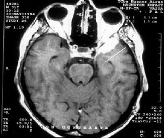
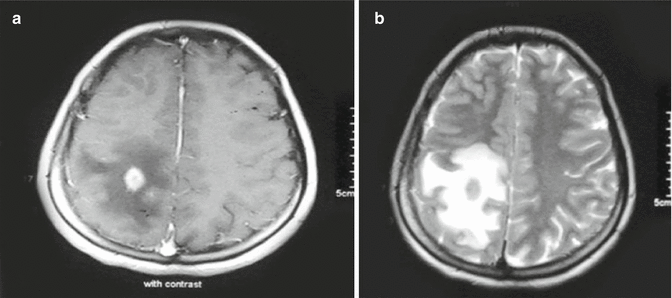
Fig. 6.2
Cerebral magnetic resonance image of a patient with neurobrucellosis due to Brucella suis. Areas of increased signal consistent with granulomas can be seen in the left parietal and temporal lobes (From Wallach et al [32], with permission)
Fig. 6.3
A nodule in the right parietal region surrounded by vasogenic edema on T1- weighted (a) and T2-weighted (b) gadolinium-enhanced axial images (From Erdem et al. [13], with permission)
6.5 Treatment
The crucial therapeutic component of all forms of human brucellosis is the administration of effective antibiotics for an adequate length of time. Importantly, the treatment of CNS complications of brucellosis may be problematic because it is needed to achieve high concentrations of drugs in the CSF.
A variety of drugs have activity against Brucella spp., though the results of in vitro susceptibility tests are not always consistent with clinical efficacy. The treatment recommended by the World Health Organization for uncomplicated brucellosis in adults is doxycycline 100 mg twice daily for 6 weeks and streptomycin 1 g/day intramuscularly administered for 2–3 weeks. Rifampicin is recommended as principal alternative therapy [8]. In clinical practice doxycycline plus rifampicin an all-oral regimen eliminates the need for parenteral administration and may allow for better compliance [3].
The paucity of therapeutic data precludes any recommendations to be offered for the time being for neurobrucellosis. Since tetracyclines and aminoglycosides do not penetrate the blood/brain barrier well, it is recommended that drugs which achieve this, such as rifampicin, trimethoprim/sulfamethoxazole (TMP/SMZ), ciprofloxacin, or ceftriaxone, be added to the standard regimen of doxycycline plus streptomycin [8, 12].
There is no data in the literature on whether brucellar meningitis or meningoencephalitis can be treated with oral antibiotics or whether an intravenous extended-spectrum cephalosporin should be added to the regimen. In various studies, ceftriaxone was found to be the most effective extended-spectrum cephalosporin for Brucella species. In addition the use of ceftriaxone alone in patients with brucellosis has been known to be associated with frequent therapeutic failures and relapses.
In a retrospective study including 215 adult patients, the first group (P1, n = 120) received ceftriaxone, rifampicin, and doxycycline [12]. The second protocol (P2, n = 44) consisted of TMP/SMZ, rifampicin, and doxycycline. In the third protocol (P3, n = 51), however, the patients started with P1 and transferred to P2 when ceftriaxone was stopped [12]. The treatment period was shorter with the ceftriaxone-based regimens (p = 0.002), and the efficacy of this regimen was found to be better when a relapse and therapeutic failure were considered [12].
The optimal duration of treatment for neurobrucellosis has not been reported, though a minimum of 6–8 weeks, and possibly longer, depending on the clinical response, has been recommended by most authorities [8]. The duration of treatment ranged from 3 months to 1 year in various clinical case studies [1, 6, 18, 33].
A total of 35 publications and 187 neurobrucellosis cases from Turkey were evaluated in a review [16]. The duration of antibiotic therapy was reported for 56 patients in the publications as 2–15 months, median 5 months [16]. In this study, ceftriaxone, doxycycline, rifampicin, streptomycin, TMP/SMZ, gentamicin, and ofloxacin were the drugs used in different combinations and over different intervals [16].
Related posts:


Stay updated, free articles. Join our Telegram channel

Full access? Get Clinical Tree


