Calcified Suprasellar Mass
Anne G. Osborn, MD, FACR
DIFFERENTIAL DIAGNOSIS
Common
Atherosclerosis, Intracranial
Craniopharyngioma
Meningioma
Aneurysm
Saccular Aneurysm
Fusiform Aneurysm, ASVD
Less Common
Neurocysticercosis
Pilocytic Astrocytoma
Dermoid Cyst
Rare but Important
Pituitary Macroadenoma
Tuberculosis
Chondroid Tumor
ESSENTIAL INFORMATION
Key Differential Diagnosis Issues
Is Ca++ curvilinear, punctate, globular, etc.?
Does lesion enhance?
Helpful Clues for Common Diagnoses
Atherosclerosis, Intracranial
Curvilinear Ca++
Usually bilateral
Often multifocal
Older patients
Craniopharyngioma
Globular, punctate, &/or ring Ca++
Younger patients (older adult tumors more often solid, Ca++ less frequent)
Meningioma
Psammomatous (sand-like) Ca++
Solid > rim enhancement
Middle-aged, older patients (unless NF2)
Aneurysm
Saccular Aneurysm
Calcification less common than with fusiform aneurysm, ASVD
Curvilinear (peripheral arcs, rings) pattern
Fusiform Aneurysm, ASVD
Linear ± rim Ca++
Ca++ often present in other vessels
Helpful Clues for Less Common Diagnoses
Neurocysticercosis
Nodular calcified stage
Usually parenchymal > > cisternal Ca++
Pilocytic Astrocytoma
Common in children, young adults
Ca++ uncommon in hypothalamic PA
Dermoid Cyst
20% have capsular Ca++
Contain lipid
Look for evidence of rupture (fatty droplets in subarachnoid spaces, cisterns)
No enhancement unless chemical meningitis
Helpful Clues for Rare Diagnoses
Only 1-2% of macroadenomas calcify
TB, healing/healed granulomatous infections cause parenchymal > > cisternal Ca++
Chondromas, enchondromas arise from central base of skull
Image Gallery
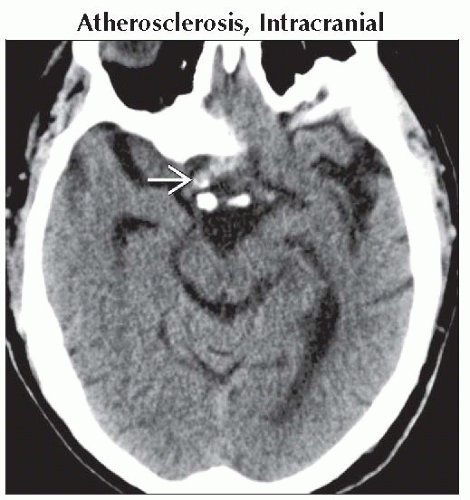 Axial NECT shows a fusiform, partially calcified mass in the suprasellar cistern
 that represents an ectatic, supraclinoid, internal carotid artery with calcified atherosclerotic plaque. that represents an ectatic, supraclinoid, internal carotid artery with calcified atherosclerotic plaque.Related posts:Stay updated, free articles. Join our Telegram channel
Full access? Get Clinical Tree
 Get Clinical Tree app for offline access

|
