Chapter 11
Case Studies Revisited
Chapter objectives
- Consolidate information presented in Chapters 6–10 regarding intervention options and their application to individuals with upper limb hypertonicity.
- Discuss the implementation and outcomes of interventions chosen for the case studies that were introduced in Chapter 5.
Abbreviations
| AROM | Active range of motion |
| BoNT-A | Botulinum neurotoxin-A |
| CMC | Carpometacarpal (joint) |
| DIP | Distal interphalangeal (joints) |
| FF | Fingers flexed |
| FE | Fingers extended |
| HIPM | Hypertonicity Intervention Planning Model |
| IF | Index finger |
| IP | Interphalangeal (joints) |
| LF | Little finger |
| MASMS | Modified Ashworth Scale of Muscle Spasticity |
| MCP | Metacarpophalangeal (joints) |
| MF | Middle finger |
| MTS | Modified Tardieu Scale of Muscle Spasticity |
| PIP | Proximal interphalangeal (joints) |
| POP | Plaster of Paris |
| PROM | Passive range of motion |
| RF | Ring finger |
| ROM | Range of motion |
| UMNS | Upper motor neuron syndrome |
11.1 Wendy—intervention process and outcomes
11.1.1 Serial and inhibitive casting
A summary of Wendy’s daily-life goals and the clinical aims associated with these goals is provided in Table 5.4. As indicated in Chapter 5 (Section 5.1.9), the primary interventions chosen were serial and inhibitive casting of the wrist and hand to:
- lengthen contractures in finger flexors, wrist flexors and thumb adductors (serial casting).
- reduce severe hypertonicity/spasticity in finger flexors (inhibitive casting).
The casting series was conducted as follows:
- Three wrist and hand casts were applied, each remaining in place for one week. Casts were made of plaster of Paris (POP) with an outer layer of fibreglass. POP was considered the most suitable casting material as the cast needed to be moulded closely to Wendy’s hand and thumb to promote a gentle curve at her fingers and to ensure that the base of her thumb and thumb webspace were well supported in partial abduction/circumduction. An outer fibreglass layer was added to provide water resistance and strength to the fingerpan.
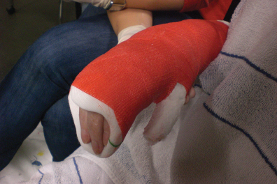
- The first cast was applied in approximately 30° of wrist flexion (Figure 11.1), which provided a submaximal stretch to Wendy’s finger flexors, since her baseline PROM for wrist extension with fingers extended (as they would be positioned in the cast) was to 20° of flexion. That is, the wrist position was “dropped back” by 10° from her maximum PROM. Care was taken to position the finger MCPs in slight flexion and mould the cast firmly under the MCPs to support their positioning. The thumb IP was positioned in neutral, with the cast fitting closely on the dorsal surface of the distal thumb phalanx to prevent hyperextension.
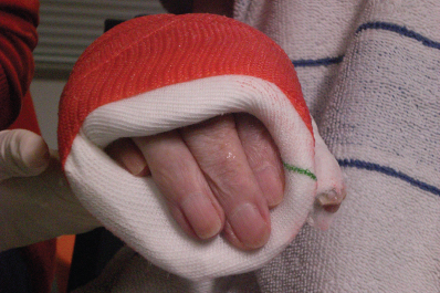
- The finger pan of the cast was cut back on the dorsum of the fingers (see Appendix 8.A) to finish just distal to the finger PIPs (Figure 11.2). The PIPs were kept enclosed in the cast to ensure that flexor digitorum superficialis (Wendy’s finger flexor muscle most affected by hypertonicity and contracture) was placed on continuous stretch by supporting the flexor surface of the joints with the finger pan while maintaining a close fit over the dorsum of the proximal phalanx of each finger. To prevent pressure areas over the PIPs, Duoderm™ was applied over the dorsum of each joint before cast application.
- Wendy reported that for the first day after the initial cast was applied, she had felt a “stretching pain” in her wrist and fingers but that this had subsided with a mild pain relief medication. By the following day the pain had resolved. Mild oedema on the dorsum of the hand was noted after removal of the second and third casts, but this resolved with the application of an elasticised stockinette and periodic elevation of the hand. Some joint stiffness was evident on passive ranging after removal of the final cast.
- Wendy had queried whether it would be necessary to support her casted arm in a sling. It was recommended that, if her shoulder became sore with the weight of the cast, she could wear a sling for some of the time. However, if no shoulder pain was evident, it was suggested that she work on isolation of shoulder and elbow movements while the cast was in place to stabilise her wrist and hand.

Figure 11.1 Wendy’s first cast was positioned in the submaximal range of approximately 30° wrist flexion.

Figure 11.2 The dorsum of the finger pan was cut back to finish just distal to the finger PIPs to keep these joints enclosed and ensure that flexor digitorum superficialis was maintained in a position that provided prolonged stretch.
Table 11.1 provides Wendy’s ROM, hypertonicity and spasticity measures after the third cast, and compares them to the baseline measures taken at initial assessment. As the cast only included Wendy’s wrist and hand, shoulder and elbow measures are not included.
Table 11.1 Range of motion, stiffness and spasticity: comparison between Wendy’s pre- and post-intervention measures.
| Right UL | AROM | PROM (R2) | MASMS | MTS (X) | Catch (R1) | Comments |
| Supination | To mid-position (no change) | 60° (no change) | 0 (↓ from 1) | 0 (↓ from 1) | No catch elicited | |
| Wrist extension (FF) | Nil (no change) | 35° (↑ by 20°) | 0 (↓ from 1+) | 0 (↓ from 1) | No catch elicited | |
| Wrist extension (FE) | Nil (no change) | 30° (↑ by 50°) | 1 (↓ from 3) | 2 (no apparent change) | 10° | Spasticity angle = R2–R1 = (30–10) = 20°a Contracture angle = (Full ‘normal’ PROM)—(‘catch’) = (70°)–(10°) = 60° |
| Finger MCP flexion | 10°–20° at IF, MF and RF during grasp (IF/MF no change; RF ↑ by 20° from hyperextension)b | Full (maintained) | Tissue stiffness (no change) | 0 (no change) | No catch elicited | Less MCP hyperextension when opening hand |
| LF PIP extension | Active movement from 90° flexion to 70° flexion when opening hand (previously positioned at 90° flexion with no active movement; increased extension at other PIPs compared to flickers only pre-casting) | To 20° of flexion (−20°) (↑ by 20°) | NA (no change) | 0 (no change) | No catch elicited | LF PIP did not have fixed joint contracture at 40° flexion |
| Thumb | ||||||
| 30° (no change) | 45° (↑ by 10°) | Nil detected (no change) | 0 (no change) | No catch elicited | Active abduction with some IP and MCP hyperextension, though observably less than pre-casting. |
| Nil (no change) | 45° (↑ by 45°) | Nil detected (no change) | 0 (no change) | No catch elicited |
a The spasticity angle on the MTS is difficult to interpret when the available passive range of motion at a joint has previously been limited by contracture and is then increased (i.e. the amount of contracture is reduced) following intervention, as illustrated in Wendy’s situation. In cases such as this it is important not to interpret an increase in spasticity angle as a failure of the intervention (for example, here the post-intervention spasticity angle of 20° is larger than the pre-intervention spasticity angle of 10°); rather, the clinician needs to consider (i) the shift in the point at which the catch becomes evident (in this case, at 10° wrist extension post-intervention compared with 30° wrist flexion pre-intervention), an improvement of 40° available range without the evidence of spasticity, and/or (ii) the difference in amount or range of ‘contracture angle’ as the measure of intervention outcome. Thus, as noted above, Wendy’s pre-intervention wrist contracture angle was 100° (measured as [70° wrist extension, which is ‘normal’ expected PROM at the wrist] + [the point of the catch, which was at 30° wrist flexion]), while her post-intervention contracture angle was 60° (measured as [70° wrist extension, ‘normal’ wrist PROM]—[the point of the catch, which was at 10° wrist extension]). Therefore, overall, the difference in contracture angle between pre- and post-intervention is 40° (that is, 100°—60°).
b Previously no active flexion at RF.
In summary, immediate outcomes from casting included:
- Reduction of contractures in Wendy’s:
- finger flexors (50° increase in PROM for wrist extension with fingers extended, from 20° wrist flexion to 30° wrist extension, see Figures 11.3 and 5.2)
- wrist flexors (20° increase in PROM for wrist extension with fingers flexed, from 15° to 35° wrist extension)
- thumb adductor (10° increase in PROM for thumb abduction, from 35° to 45° abduction)
- extensor pollicis longus (45° increase in PROM for thumb IP flexion, from 0° at initial assessment)
- little finger PIP joint (20° increase in PROM for PIP extension, from 40° to 20° flexion, indicating that initial joint limitation was at least partially due to joint stiffness rather than joint contracture).
- finger flexors (50° increase in PROM for wrist extension with fingers extended, from 20° wrist flexion to 30° wrist extension, see Figures 11.3 and 5.2)
- Reduction of hypertonicity (stiffness) in:
- pronators (MASMS reduced from 1 to 0)
- wrist flexors (MASMS reduced from 1+ to 0)
- finger flexors (MASMS reduced from 3 to 1)
- pronators (MASMS reduced from 1 to 0)
- Reduction in spasticity angle in finger flexors (see Table 11.1 footnote)
- When attempting to grasp an object, Wendy was now able to achieve more extension of her fingers from the PIPs and DIPs (at least 20° and up to 60° joint excursion at each finger joint compared with only flickers of movement pre-casting). While slight MCP hyperextension was still apparent with finger extension, this was reduced compared to pre-casting. She could still only actively abduct her thumb to 30°, but the slight increase in passive thumb abduction made it easier for her to place the item into her hand and she used a combined adduction/partial opposition movement to place her thumb around it. Wendy’s thumb IP remained in neutral to slight hyperextension. When grasping, slight active MCP flexion was apparent at all fingers except the little finger, together with less extreme flexion at the PIPs and DIPs

Stay updated, free articles. Join our Telegram channel

Full access? Get Clinical Tree


