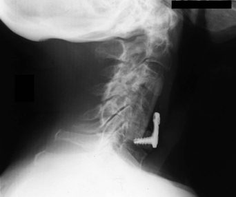
31 A 79-year-old woman sustained central cord syndrome after a fall, with significant upper extremity weakness and incontinence. She had multilevel cervical disc hernia-tions and concomitant osteoporosis. Magnetic resonance imaging (MRI) showed cervical spondylosis with a large soft herniated disc at C4–C5 and cord signal change. Due to progressive neurologic deterioration, the decision was made to treat her surgically. Postoperative lateral cervical x-ray displays a fibular strut graft spanning from C3 to C6 with a buttress plate and significant osteopenia (Fig. 31-1). Central cord syndrome with acute disc An anterior cervical discectomy at C3–C4 and C5–C6, with C4 and C5 corpectomies, and reconstruction with a fibular strut graft, arthrodesis, and buttress plate were completed. Central cord syndrome is a partial neurologic deficit, with motor more than sensory loss, upper more than lower extremity deficit, and distal more than proximal involvement. Pathologically, one sees a significant cord contusion. Acute decompression has been thought to carry a higher neurologic morbidity, and surgery is usually performed on a delayed, elective basis. If signs of progressive neurologic deterioration are apparent, then acute decompression is warranted. Also, if the patient has an acute disc herniation or findings suggesting instability, such as dislocation, more urgent surgical intervention may be warranted. This patient had the neurologic indications for surgery but carried a high risk of nonunion due to her age, osteoporosis, and multilevel fusion. She wore a cervical collar for 3 monthsafter surgery and was given a cervical bone stimulator. Her recovery went well, with resolution of the incontinence and improvement of her upper extremity strength.
Central Cord with Osteoporosis
Presentation
Radiologic Findings
Diagnosis
Treatment
Discussion
Central Cord with Osteoporosis
Only gold members can continue reading. Log In or Register to continue


Full access? Get Clinical Tree


