Central Sleep Apnea


Central Sleep Apnea


Richard B. Berry


OVERVIEW

Cessation of breathing during sleep is referred to as apnea (in adults, >10 seconds by convention), whereas reductions in breathing are called hypopneas (1,2). Cessation of breathing during sleep despite ongoing respiratory effort is associated with obstruction of the upper airway (obstructive apnea), whereas the loss of ventilatory effort can also lead to the loss of airflow (central apnea) (3). The term central sleep apnea (CSA) is used to describe both the events and the clinical disorders characterized by repeated cessations of airflow during sleep, resulting from temporary loss of ventilatory effort (Fig. 23-1) (4). Although this chapter is focused on CSA, we acknowledge that central apneas are uncommonly seen in isolation. The majority of patients with central apneas will also have some obstructive apnea and vice versa (5). By extension, it follows that the mechanisms responsible for the different types of apnea must overlap (6). A diagnosis of the CSA syndrome is usually made when the majority of events are central in nature (arbitrarily >≈50%). As will be discussed, the CSA syndromes are a heterogeneous group of disorders that can be broadly classified on the basis of arterial carbon dioxide levels during wakefulness (hypercapnic vs. eucapnic/hypocapnic; see Table 23-1).


VENTILATORY CONTROL AND PATHOPHYSIOLOGIC MECHANISMS


Ventilatory Control

Control of ventilation is dependent on both metabolic (automatic, i.e., chemoreceptors, stretch receptors) and behavioral (wake-dependent) control systems (7). During NREM sleep, ventilation is critically dependent on the metabolic control system. Under metabolic control, ventilation can be stimulated through medullary brain stem centers by CO2 (likely via H+ concentration) and the carotid body by PO2 and PCO2. During wakefulness, an additional nonspecific “wakefulness drive” maintains ventilation even if the PCO2 is reduced below the eucapnic level. With the onset of sleep, the wakefulness drive is lost and other factors, such as increased upper airway resistance, contribute to a normal rise in PCO2 of 4 to 6 mm Hg above the wakefulness eucapnic level (8,9). In addition, if the PCO2 falls below a certain level (the apneic threshold) during NREM sleep, ventilatory effort ceases (10). The apneic threshold is typically 2 to 6 mm Hg below the sleeping eucapnic PaCO2 level and usually corresponds roughly to the awake eucapnic PaCO2 level or slightly lower (11). This apnea threshold may, in part, explain the dysrhythmic breathing frequently seen at sleep onset (see below, sleep transition central apneas), even in normal individuals. Once a stable sleep stage is reached, ventilation should become regular under stable metabolic control.


Ventilatory Control in Central Sleep Apnea

Since the CSA syndromes are heterogeneous, the pathophysiology is also quite varied (Table 23-2). In some patients with hypercapnic CSA, ventilatory control is abnormal. These individuals have low or absent hypoxic/hypercapnic responsiveness. Respiration during wakefulness is maintained by behavioral/wakeful stimuli plus automatic mechanisms. However, during sleep, when these wakefulness-dependent mechanisms are no longer operative, there is little residual drive to ventilate because metabolic drive is attenuated. As a result, hypoventilation and central apneas frequently ensue (4).

Based on the comments above, the slope of the ventilatory response to hypercapnia (frequently measured
during wakefulness) may be an important variable in the development of central apneas during sleep (12). In patients with markedly diminished or absent chemosensitivity, some form of sleep-disordered breathing (SDB), frequently central apnea, would be expected. If carbon dioxide sensitivity is very low or absent, there would be little ventilation stimulus during sleep and central apneas with sustained hypoventilation would be predictable. Patients with disorders such as central alveolar hypoventilation (“Ondine’s curse”) and the obesity-hypoventilation (Pickwickian) syndrome would fall into this category (13,14 and 15). Similarly, recent reports have suggested a high prevalence of CSA among patients chronically taking narcotic medications (which can inhibit chemosensitivity) (16).


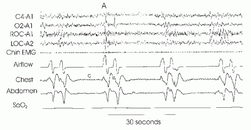
FIGURE 23-1 Idiopathic central apnea. The figure illustrates three apneas (cessation of airflow) without respiratory effort detectable in the chest and abdominal belts. Termination of the first apnea is associated with arousal (A). The small deflections in the chest tracing during apnea (c) do not reflect respiratory effort but are secondary to cardiac pulsations.


TABLE 23-1 CLASSIFICATION OF CSA SYNDROMES


HYPERCAPNIC CSA


1.


Central hypoventilation


a.


Idiopathic central hypoventilation (Ondine’s curse)


b.


Brain tumors


c.


Cerebrovascular disease


d.


Chronic narcotic use


e.


Obesity hypoventilation syndrome


2.


Neuromuscular disorders


a.


Myasthenia gravis (neuromuscular junction)


b.


ALS (motor neuron disease)


c.


Post poliosyndrome


d.


Myopathies (e.g., acid maltase deficiency)


3.


Chest wall syndromes (kyphoscoliosis)


NONHYPERCAPNIC CSA


1.


Idiopathic


2.


Treatment emergent


3.


Sleep transition


4.


Cheyne-Stokes breathing


a.


CHF


b.


Neurologic disorders


5.


Periodic breathing at high altitude


On the other hand, increased hypercapnic sensitivity can also lead to ventilatory instability during sleep. These “high-drive” CSA patients often have low arterial PCO2
levels during wakefulness and a high hypercapnic ventilatory response (17). The unusually steep hypercapnic responsiveness can lead to respiratory instability with a fluctuating pattern of ventilation in the wake—sleep transition. This instability is believed to be secondary to intermittent overventilation (yielding hypocapnia) alternating with underventilation (yielding hypercapnia). The cyclic nature of ventilation probably results from the marked fluctuations in ventilation that occur with modest changes in PaCO2. Thus, elevated chemosensitivity contributes to cessations in airflow by producing ventilatory overshoots during which PaCO2 falls below the apnea threshold.


TABLE 23-2 PATHOPHYSIOLOGICAL MECHANISMS OF CSA


HYPERCAPNIC CSA


A.


“Won’t breathe”—low or absent ventilatory response to PCO2 and/or PO2


Abnormal ventilatory control centers


Abnormal chemoreceptors


Abnormal communication between chemoreceptors and control centers


Brainstem defects


B.


“Can’t breathe”


Motorneuron defects (brainstem/spinal cord)


Motor nerve lesions


Neuromuscular junction abnormalities


Myopathy


Increased respiratory impedance (chest wall disease, obesity)


EUCAPNIC/HYPOCAPNIC CSA (ULTIMATE MECHANISM: PCO2 IS BELOW THE APNEIC THRESHOLD)


A.


Idiopathic central sleep apnea


High ventilatory drive (ventilatory response to PCO2 or PCO2)


Small difference between the apneic threshold and sleeping eucapnic PCO2


High plant gain


Long transition between wake and stable sleep


B.


CSRs


High ventilatory drive (ventilatory response to PCO2 or PO2)


Small difference between the apneic threshold and sleeping eucapnic PCO2


Long circulation time (delay in ABG information to controllers)


Increase in reflex stimulation of breathing


(J receptors)—pulmonary congestion


The term “loop gain” is used to describe the net stability of the ventilatory control system (18,19,20,21,22,23,24 and 25). A high loop gain refers to one that is intrinsically unstable, whereas a low loop gain implies intrinsic stability. Loop gain is defined as the ratio of the response to the stimulus (ventilatory response/ventilatory disturbance) in a feedback control system (i.e., a large response to a minor perturbation is destabilizing). Thus, an intrinsically stable system would experience a minor response to a perturbation and continue relatively stably thereafter. On the other hand, an intrinsically unstable system may have a large response to a minor perturbation, yielding subsequent instability. In the case of breathing, a robust ventilatory response to a trivial increase in PaCO2 would be destabilizing to the control system. This loop gain concept is useful in determining the various factors that can contribute to the development of CSA.

A unifying phenomenon in all forms of CSA is a complete loss of ventilatory drive. This cessation can be recognized by an absence of electromyographic activity in the respiratory muscles or an absence of pleural pressure swings (estimated using esophageal pressure). In nonhypercapnic CSA, the basic mechanism underlying central apnea is that the PaCO2 falls below the apneic threshold (26). Following an arousal, which typically increases ventilation, apnea can occur if sleep resumes before the PCO2 rises above the apneic threshold (i.e., sleep transition apneas) or if there is a ventilatory overshoot due to control instability (i.e., high loop gain). The ventilatory response to arousal may indeed be a mechanism driving unstable ventilation in those patients with an unstable state (i.e., wake to sleep to wake transitions) (27). As an individual changes from wakefulness to stage 1 or 2 sleep, the PCO2 level that was adequate to stimulate ventilation during wakefulness may be inadequate to do so during sleep, and an apnea may occur. This apnea may arouse the individual, and the process repeats itself over and over. Thus, there is ventilatory instability with oscillating levels of ventilatory drive. The causes of this instability include high controller gain (hypercapnic sensitivity and responsiveness), high plant gain (efficient CO2 excretion), delay in information reaching the controllers (long circulation time, i.e., mixing gain), and a PaCO2 level close to the apnea threshold (11,28,29). The overall ventilatory loop gain can therefore be characterized by the product of the individual gains (i.e., controller, plant, and mixing) (30).


Pathophysiology of Cheyne-Stokes Respiration

The most common type of nonhypercapnic CSA is called Cheyne—Stokes respiration (CSR) (Fig. 23-2) (31). This disorder is characterized by a waxing and waning pattern of breathing most commonly seen in individuals with congestive heart failure (CHF) and left ventricular systolic dysfunction (32). This type of breathing is quite distinct, with a crescendo—decrescendo ventilatory pattern with a central apnea or hypopnea at the nadir. Thus, it is somewhat different from the aforementioned central apnea, which has a more abrupt onset and offset. Typically, the ventilatory phase between central apneas is longer in CSR than in other types of repetitive central apneas because of a prolonged circulation time (33). Another difference is that arousal tends to occur at the peak of the ventilatory effort in CSR rather than at event termination (34). The etiology of this breathing pattern in CSR has never been fully understood but is probably a product of respiratory control system instability (high loop gain with out-of-phase signal at the chemoreceptor) resulting from a prolonged circulation time and increased ventilatory responsiveness to rising PCO2. In an anesthetized animal, lengthening normal circulation time can induce Cheyne—Stokes breathing (CSR) (35). Such an increased circulation time may produce unstable ventilation due to the delay that occurs between mechanical changes in respiration (hyperpnea or hypopnea) and receptor stimulation resulting from changes in arterial blood gases. If a patient hypoventilates or has an apnea, a substantial period of time will pass before increasing PCO2 or decreasing PO2 is detected at the appropriate receptor. As a result, the apnea or hypopnea is prolonged. When the deoxygenated, hypercapnic blood does reach the receptor, ventilation is stimulated for a disproportionately long period of time because the corrected blood gases are again not presented to the receptor for an extended period of time. Thus, ventilation can wax and wane, with actual apneas occurring at the nadir of this cycle. Whether this is the mechanism of Cheyne—Stokes ventilation (CSR) seen with CHF is speculative, because a severalfold increase in circulation time is necessary to produce such breathing dysrhythmias in animals (36). This is probably a greater change than commonly occurs with heart failure. When carefully matched groups of CHF patients with and without CSR have been compared, the groups do not systematically differ in circulation time. On the other hand, patients with CSR in CHF do have high ventilatory drive based on increased chemosensitivity and stimulation of ventilation through pulmonary stretch receptors (37). Thus, the group with CSR does generally have a lower awake arterial PCO2 than CHF patients without CSR. One cause of the higher drive appears to be a higher wedge pressure (pulmonary arterial occlusion pressure) (38). Presumably, pulmonary congestion results in
the stimulation of J receptors in the lung, which also increases ventilatory drive. Thus, a prolonged circulation time probably must be combined with an increased ventilatory drive to yield (CSR) (39). Patients with CHF and CSR tend to maintain their waking eupneic PCO2 levels during sleep, thus keeping the PaCO2 close to the apnea threshold. Therefore, rather minor overshoots in ventilation could dramatically reduce ventilatory drive (40,41). These mechanisms (circulation time, CO2 responsiveness, CO2 set point) explain much of the variability in CSR occurrence in heart failure patients.


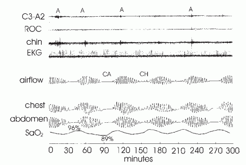
FIGURE 23-2 Cheyne-Stokes respiration. The figure illustrates a waxing and waning pattern of airflow and respiratory effort over the course of this 5-minute recording. Central apnea (CA) or central hypopnea (CH) occur at the nadirs in respiratory effort. Of note, the duration of each cycle is roughly 1 minute. The periods of ventilation between events are much longer than in idiopathic central apnea (Fig. 23-1). In addition, note the fall in oxygen saturation that occurs following each apnea. The time duration from the end of apnea to the nadir in arterial oxygen saturation (Sao2)-desaturation is prolonged and reflects circulatory delay. Although difficult to see at this resolution, there is frequently arousal (A) from sleep at the peak of the hyperpnea with associated tachycardia. In idiopathic central apnea, arousal usually occurs at apnea termination (Fig. 23-1).

CSR has also been reported in patients with neurologic disease, primarily cerebrovascular disorders. However, the actual ventilatory pattern in these patients has been less well characterized than in patients with CHF, and the mechanisms remain poorly understood. In many cases, a cardioembolic etiology for the cerebrovascular event has not been carefully excluded, leading many to suspect underlying cardiac dysfunction in such patients. In addition, fluctuations in the level of consciousness in stroke patients have been associated with central apneas (state instability), similar in mechanism to the sleep-transition apneas described earlier. Other mechanisms leading to increased ventilatory drive have also been proposed to explain CSR in stroke patients. A reduction in or loss of tonic inhibition of ventilation could lead to this instability but has not been fully characterized. Thus, the major mechanisms of CSRs in cerebrovascular disease remain to be elucidated (4).


Pathophysiology of Hypercapnic Central


Sleep Apnea—Effects of Sleep Stage

As noted above, some hypercapnic CSA patients have a defect in central drive. This may be caused by abnormal chemoreceptors (the sensors detecting PCO2 [or H+] and PO2), poor communication of this information to the controllers, or a defect in the generation of ventilatory rhythm. In some of these patients, the wakefulness drive is sufficient to maintain ventilation during wakefulness, but hypoventilation and/or central apnea occurs during sleep (8). In other patients with hypercapnic CSA, the central drive is intact but other abnormalities compromise ventilation during sleep. These include defects in the neural motor circuits (e.g., phrenic nerve injury following cardiac surgery), respiratory muscles (e.g., acid maltase deficiency), or the chest wall (e.g., kyphoscoliosis). High ventilatory drive and accessory respiratory muscle activation may partially compensate for these problems during wakefulness, but the loss of the wakefulness drive with sleep onset may result in the worsening of hypoventilation and/or frank central apnea. Of note, the majority of the patients with hypercapnic CSA have the most severe hypoventilation during rapid eye movement (REM) sleep. During this sleep stage, ventilation is under both metabolic and nonmetabolic control and there is generalized skeletal muscle hypotonia (42). Those patients with compromised respiratory muscles or chest wall abnormalities are frequently dependent on the accessory muscles of
respiration to maintain ventilation. When these muscles develop the atonia characteristic of REM sleep, the diaphragm alone may be unable to maintain adequate ventilation. On the other hand, there does exist the rare patient with central defects in ventilatory control who experiences improvements in respiration during REM sleep, as compared with NREM sleep. This phenomenon is thought to result from the nonmetabolic influences on ventilation that occur in REM sleep (e.g., dream content) but not NREM sleep. However, this REM-related improvement is relatively uncommon.


DEFINITIONS AND CLASSIFICATION OF CENTRAL SLEEP APNEA SYNDROMES

Patients with predominantly CSA constitute fewer than 10% of apneic individuals in most sleep laboratory populations, with some recent data suggesting only about 4%. As a result, only a small number of studies with more than a few such patients have been reported, which makes knowledge of this disorder limited. Most of this chapter is dedicated to a discussion of patients with CSA who breathe normally during the day. However, any patient with hypoventilation during wakefulness will almost certainly have further hypoventilation (with central apneas) at night. As stated, CSA is a heterogeneous disorder that can be classified into the hypercapnic and nonhypercapnic types (Table 23-1).


HYPERCAPNIC CENTRAL SLEEP APNEAS AND SLEEP HYPOVENTILATION SYNDROMES

Patients with hypercapnic CSA usually have daytime hypercapnia that worsens with sleep. Some authors have used the term “sleep hypoventilation syndrome” to highlight the fact that most such patients hypoventilate during sleep but actually have few discrete central apneas. These hypercapnic patients can be further separated into a “won’t breathe” group with defects in central drive but intact motor nerves, spinal cord, respiratory muscles, and lungs and a “can’t breathe” group with normal drive but defects distal to the ventilatory control centers (Table 23-2). The former group comprises the central hypoventilation syndromes. Although uncommon, one form of this syndrome can present in infancy (congenital central hypoventilation syndrome [CCHS]). With the availability of genetic testing for the Phox2b gene, there is increasing appreciation for CCHS, including some cases presenting in adulthood (13). In others, the neural pathways from these medullary respiratory neurons to the motorneurons of the ventilatory muscles are interrupted, which may occur after cervical cordotomy. Because the brainstem is the primary source of both ventilatory pattern generation and the processing of respiratory afferent input from chemoreceptors and intrapulmonary receptors, any disease process affecting this area could influence ventilation during sleep. Damage to the brainstem, particularly the medullary area, may lead to hypoventilation during wakefulness but more commonly affects ventilation during sleep. Other processes, such as tumor, infarction, hemorrhage, Arnold Chiari malformation, or encephalitis, can affect the medullary area, leading to breathing dysrhythmias during sleep, with central apneas being a prominent feature (43).

The second group of hypoventilation syndromes (unimpaired drive—“can’t breathe”) are commonly classified anatomically, including lesions of the upper motor neurons (e.g., amyotrophic lateral sclerosis [ALS], which can involve both upper and lower motor neurons), the spinal cord (e.g., trauma), the anterior horn cells (e.g., poliomyelitis), the lower motor neurons (e.g., phrenic nerve C3-C5), the neuromuscular junction (e.g., myasthenia gravis), and the respiratory muscles themselves (e.g., polymyositis, acid maltase deficiency). Mechanical impairment of the chest wall (e.g., kyphoscoliosis) and lung parenchyma (e.g., chronic obstructive pulmonary disease) can also lead to hypercapnic respiratory failure. Although these disorders may be characterized by central apneas during sleep, the principal problem is nocturnal hypoventilation (44), particularly during REM sleep, which leads to substantial hypoxemia and hypercapnia with their associated sequelae. Thus, the hypercapnic CSA syndromes include a spectrum of disease from hypoventilation to frank cessation in breathing (45,46).

Finally, as stated previously, any neurologic disorder affecting the ventilatory control system could influence ventilatory patterns during sleep, possibly leading to central apneas. Thus, patients with autonomic dysfunction, such as the Shy—Drager syndrome, familial dysautonomia, or diabetes mellitus, may have central apneas, although the precise mechanisms leading to such apneas have not been determined.


NONHYPERCAPNIC CENTRAL SLEEP APNEA

Patients with nonhypercapnic CSA have normal or low daytime PCO2. Syndromes falling under this category include idiopathic CSA and CSA with CSR (Table 23-1). We have also included sleep-transition CSA and treatment-emergent CSA for completeness, although these are really forms of isolated CSA rather than true CSA syndromes (Figs. 23-3 and 23-4). Sleep-transition central apneas may occur in normal individuals at sleep onset, when there is a waxing and waning of ventilation (47). These sleep-transition apneas occur when there is state instability (transitions from sleep to wake), with concomitant fluctuations in PCO2. Therefore, any process that leads to frequent sleep—wake transitions over the course of the night (e.g., insomnia, periodic limb movements, obstructive
apneas, spontaneous arousals) may increase the number of central apneas. Treatment-emergent central apneas (also referred to as complex apnea (48,49 and 50)) occur during the titration of continuous positive airway pressure (CPAP) in obstructive sleep apnea (OSA) patients (Fig. 23-4). Although this has been studied minimally and the explanation remains obscure, CPAP does lower upper airway resistance, which can lead to falls in PCO2. If the PCO2 falls below the apnea threshold, central apneas will ensue (51). Increased lung volume on CPAP may also activate stretch receptors that can inhibit ventilation. Clinical experience and emerging data suggest that this form of central apnea generally resolves with ongoing therapy. A similar phenomenon has been observed in OSA patients following tracheostomy, wherein some individuals will experience central apneas that eventually resolve (52). Patients with idiopathic CSA have no other identifiable disorder (i.e., diagnosis of exclusion). However, this syndrome is relatively rare (53).

Only gold members can continue reading. Log In or Register to continue

Stay updated, free articles. Join our Telegram channel

Jul 14, 2016 | Posted by in PSYCHIATRY | Comments Off on Central Sleep Apnea

Full access? Get Clinical Tree

Get Clinical Tree app for offline access