•Central sleep apnea with Cheyne-Stokes breathing
•Central apnea due to a medical disorder without Cheyne-Stokes breathing
•Central sleep apnea due to high altitude periodic breathing
•Central sleep apnea due to a medication or substance
•Primary central sleep apnea
•Primary central sleep apnea of infancy
•Primary central sleep apnea of prematurity
•Treatment-emergent central sleep apnea
Positional Dependency in Central Sleep Apnea Due to Hyperventilation Not Related to Chronic Heart Failure
Clinical Presentation
In CSA due to hyperventilation not related to chronic heart failure, positional dependency has been reported in only a few cases.
Zaharna et al. [2] recently described a 29-year-old male with severe idiopathic central sleep apnea with an apnea-hypopnea index (AHI) of 49.3 per hour and a central apnea-hypopnea index (CAHI) of 42.7 per hour. Positional dependency was present with a supine CAHI of 101.6 per hour in contrast to a right lateral CAHI of 7.1 per hour and left lateral CAHI of 39 per hour. The patient denied any symptoms of an underlying cardiac, renal or neurological condition, and no further diagnostic tests were performed. He was successfully treated with CPAP and he declined positional therapy.
Another case of positional CSA was presented by DelRosso et al. [3]. A 66-year-old man with hypertension complained of excessive daytime sleepiness, dyspnoea upon exertion, episodes of dream-enacting behaviour and tongue biting when sleeping in the right lateral decubitus position. Polysomnography (PSG) revealed severe positional CSA, with predominance in the supine position (187 events) and right lateral decubitus position (35 events), and only 2 events in the left lateral decubitus position. Obstructive events were minimal (6 events). MRI scan of the brain revealed a calcified left vertebral artery that caused compression of the pre-Bötzinger complex in the left anterolateral medulla and patient was treated with BIPAP-ST.
Oksenberg et al. [4] presented a 57-year-old female with both obstructive and central sleep apnea after an ischemic cerebrovascular accident. In this patient the first PSG was performed 2 months post-stroke and revealed severe CSA/CSB in non-REM and a severe OSA pattern during REM sleep, independent of body position (RDI supine 85.2 per hour and lateral 95.4 per hour). After 3 months and overall clinical improvement, a second PSG showed a complete disappearance of breathing abnormalities in the lateral position for both CSB and OSA (RDI 0 per hour) but only a small improvement in supine position (supine RDI 73.2 per hour), with the CSA/CSB characterized mainly by central hypopneas. Finally, Yaegashi et al. [5] reported a statistically significantly higher total CAHI in supine position during NREM sleep in complex sleep apnea syndrome during the baseline polysomnography, but these data can hardly be considered clinically relevant (2.5 ± 3.1 vs. 0.9 ± 2.3).
Possible Mechanisms of Positional Central Sleep Apnea Due to Hyperventilation Not Related to Chronic Heart Failure
Development of central sleep apnea in the supine position, although unexpected, can be explained by a considerable overlap in the pathogenesis and pathophysiology of central and obstructive apneas [6–8]. The behaviour of the upper airway in the case of obstruction may not be uniform. Badr et al. [9] described the occurrence of gradual progressive pharyngeal narrowing during induced hypocapnic central apnea and frequent complete pharyngeal occlusion in 146 of 160 investigated cases. Resumption of inspiratory effort was associated with persistent narrowing or complete occlusion unless electroencephalographic arousal was noted. They concluded that subatmospheric intraluminal pressure is not required for pharyngeal occlusion to occur and may be due to passive collapse or active constriction. Vanderveken et al. [10] reported that during central apnea, respiratory impedance was highly variable ranging from a wide open pharynx to complete upper airway closure. In 20 % of analysed central events, a definite closure of the upper airway occurred as demonstrated by Zrs values of 35–57 hPa/l/s. In 10 % an increase by >200 % Zrs was observed, reaching a value of 22–23 hPa/l/s. In 70 %, only a partial occlusion or no change at all in airway patency occurred with Zrs < 17 hPa/l/s. Pepin et al. [11] reported on a case of central apnea with upper airway collapse visualized by somnofluoroscopy. In this patient, the pharyngeal collapse occurred at the end of the central events. On the other hand, Guilleminault et al. [12] did not find evidence for complete upper airway collapse during central apnea in an EMG study. Another argument for upper airway closure during central events is the effectiveness of CPAP for the treatment of central apnea, with a role for the upper airway factor in the pathogenesis of central sleep apnea [13, 14].
Altogether, the aforementioned obstructive component reported in central apnea could explain the occurrence of supine dependency in some patients. It is however in contradiction with the traditional paradigm that central apnea is characterized by recurrent apnoeic episodes in the absence of upper airway obstruction during sleep, according to the guidelines of the AASM [15].
Airway obstruction, especially in the supine position, can mimic central apnea, and nearly 30 % of obstructive events appeared phenomenologically as central apneas in a study by Luo et al. [16]. On the other hand, central hypocapnic hypopnea may exhibit obstructive features [8]. Boudewyns et al. [17] pointed at methodological issues in the assessment of central apnea, with an overestimation of central apneas based on strain gauges in patients with obstructive sleep apnea syndrome. In this perspective, supine dependency in misclassified obstructive events is obvious.
Positional Dependency in Central Sleep Apnea Due to Hyperventilation, Type CSA/CSB Related to Chronic Heart Failure
CSA/CSB in Heart Failure Patients: Clinical Picture and Pathophysiology
CSA/CSB is common in chronic heart failure patients with reported prevalences of 30–40 % [18, 19] and is also observed in stroke patients and in patients with renal failure. CSA/CSB is characterized by a typical waxing and waning breathing pattern, with a relatively long cycle length of about 60 s (Fig. 1). The pathophysiology underlying this typical breathing pattern is complex, and detailed discussion is beyond the scope of this section (please refer to Yumino and Bradley [20] for further reading). Briefly, CSA/CSB results from activation of pulmonary stretch receptors due to pulmonary congestion and enhanced sensitivity of the peripheral and central chemoreceptors leading to hyperventilation and intermittent ventilatory overshooting. This mechanism cyclically drives the PaCO2 below the CO2 apnoeic threshold, and the ventilatory instability is further enhanced by increased circulation time in heart failure, hypoxaemia, arousals causing sleep stage shifts and sleep state instability with further aggravation of CSA/CSB [21], metabolic alkalosis and upper airway narrowing (Fig. 2).
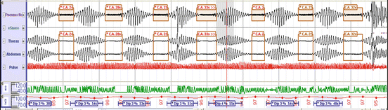
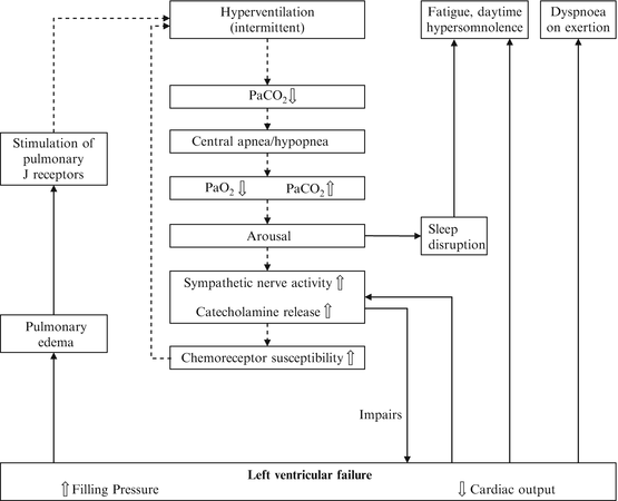
Fig. 1
PSG recording of central sleep apnea type Cheyne-Stokes breathing
Fig. 2
Mechanisms involved in the pathophysiology of central sleep apnea type Cheyne-Stokes breathing. Reprinted with permission from the Neth Heart J. 2010 May;18(5):260-3
CSA/CSB is present predominantly in sleep stages N1 and N2, much less in slow wave sleep and usually absent in REM sleep [22], supposedly because the atonia of REM sleep is ‘protective’ against the hyperventilation in CSA/CSB and the absence of the CO2 threshold in REM sleep. Moreover, it is more predominant at the end of the night. This may be due to the prevalence in sleep stages N1 and N2 on the one hand but on the other hand also may reflect a gradual fluid shift from the extravascular peripheral space rostrally to the lungs, thereby worsening pulmonary congestion [23]. Orthopnoea is one of the nocturnal signs of heart failure, and patients report relief when sleeping with their upper body in an elevated angle.
Positional CSA/CSB: Prevalence, Severity and Clinical Determinants
We performed a literature search using PubMed as well as Web of Science, using the keywords ‘central sleep apnea’, ‘CSA’, ‘Cheyne Stokes breathing’ and ‘CSB’, combined with ‘position dependent’, ‘position’, ‘positionality’, ‘lateral’ or ‘supine’. Only studies reporting on systematically obtained, original data were included.
Sahlin et al. [22] reported on 20 consecutive patients with CSA/CSB of which 18 had congestive heart failure (NYHA classes II–III) and 2 had atrial fibrillation and experienced a stroke before. The mean central AHI (CAHI) was 31 ± 9 per hour, and the obstructive AHI was 2 ± 2 per hour. The average BMI was 27 ± 3 kg/m2 and only two subjects reported snoring. The CAHI was higher in supine as compared with the non-supine position in 17 out of 20 patients (mean supine CAHI 41 ± 13 and non-supine CAHI 26 ± 12, p < 0.001), and BMI did not correlate with the postural effect of central sleep apnea.
Szollosi et al. [24] also reported on 20 patients (age 59.9 ± 2.3 years) with stable heart failure (mean left ventricular ejection fraction 26.5 ± 2.2 %) and CSA/CSB with an average BMI of 26.5 ± 0.8 kg/m2. Total AHI was 26.4 ± 3 per hour, with 21.2 ± 2.7 central, 3.3 ± 1.6 mixed and 2.1 ± 0.4 obstructive events per hour. The lateral body position was associated with a significant reduction in AHI in all sleep stages: stage N1 54.7 ± 4.2 per hour vs. 27.2 ± 4.1 per hour, stage N2 43.3 ± 6.1 per hour vs. 14.4 ± 3.6 per hour, SWS 15.9 ± 6.4 per hour vs. 5.4 ± 2.9 per hour and REM 38.7 ± 7.3 per hour vs. 11.0 ± 3.0 per hour (p for all <0.01). In addition, the lateral position was associated with reduced AHI-related hypoxemia during sleep stages 1 and 2, during which most events occurred, but event duration was not influenced by sleep stage nor sleeping position.
Joho et al. [25] found that 12 (48 %) of 25 patients with stable heart failure (left ventricular ejection fraction <0.45, a mean AHI 28 ± 8 per hour and CAHI 21 ± 10 per hour) had positional CSA. Overall, subjects with positional CSA/CSB had a lower AHI, a lower NYHA score and better physical activity; used β-blockers; had a lower BNP (brain natriuretic peptide); and a better LVEF. When eight patients with non-positional CSA/CSB were intensively treated to improve cardiac function, non-positional CSA/CSB changed to positional CSA/CSB in all eight patients.
In our own sleep laboratory in a tertiary referral centre, we looked at 28 consecutive patients with CSA/CSB (27 males, mean age 66.3 ± 10 years , mean BMI 30.1 ± 5.1 kg/m2, mean AHI 42 ± 19.7, with ≥30 min in either supine and non-supine sleeping position, and >50 % central events type CSB). Nineteen patients used PAP therapy during the sleep registration. Mean AHI in non-supine position was 34.5 ± 20.3 vs. AHI supine 55.0 ± 23.9 (p < 0.001). In 25 patients AHI supine was higher than AHI non-supine. Positional CSA, defined as an AHI ratio non-supine/supine ≤0.5, was seen in 11 out of 28 patients (39 %). See Fig. 3 for an example of positional CSA/CSB in a young male patient with atrial fibrillation. In positional CSA average, AHI was 29 ± 16.7, while in non-positional CSA, average AHI was 50.4 ± 16.9 (p = 0.003). If positional CSA was present, average AHI non-supine was 15.9 ± 11.8 as compared with AHI supine 49.3 ± 24.3 (p = 0.003; unpublished data).
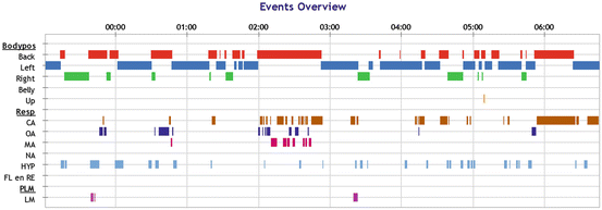
Fig. 3


Positional CSA/CSB. Red bar: supine position; green bar: right lateral decubitus position; blue: left lateral decubitus position. CA central apnea, OA obstructive apnea, MA mixed apnea. Central apneas were of Cheyne-Stokes breathing type. Most central events cluster with the supine body position
Related posts:


Stay updated, free articles. Join our Telegram channel
Full access? Get Clinical Tree
