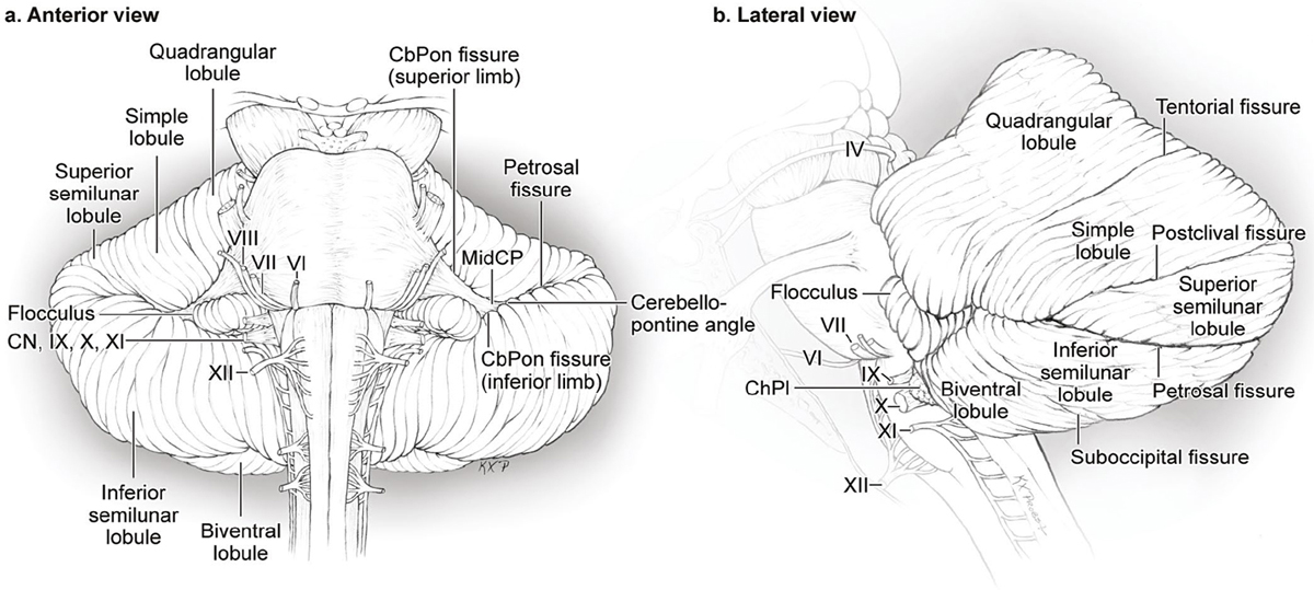
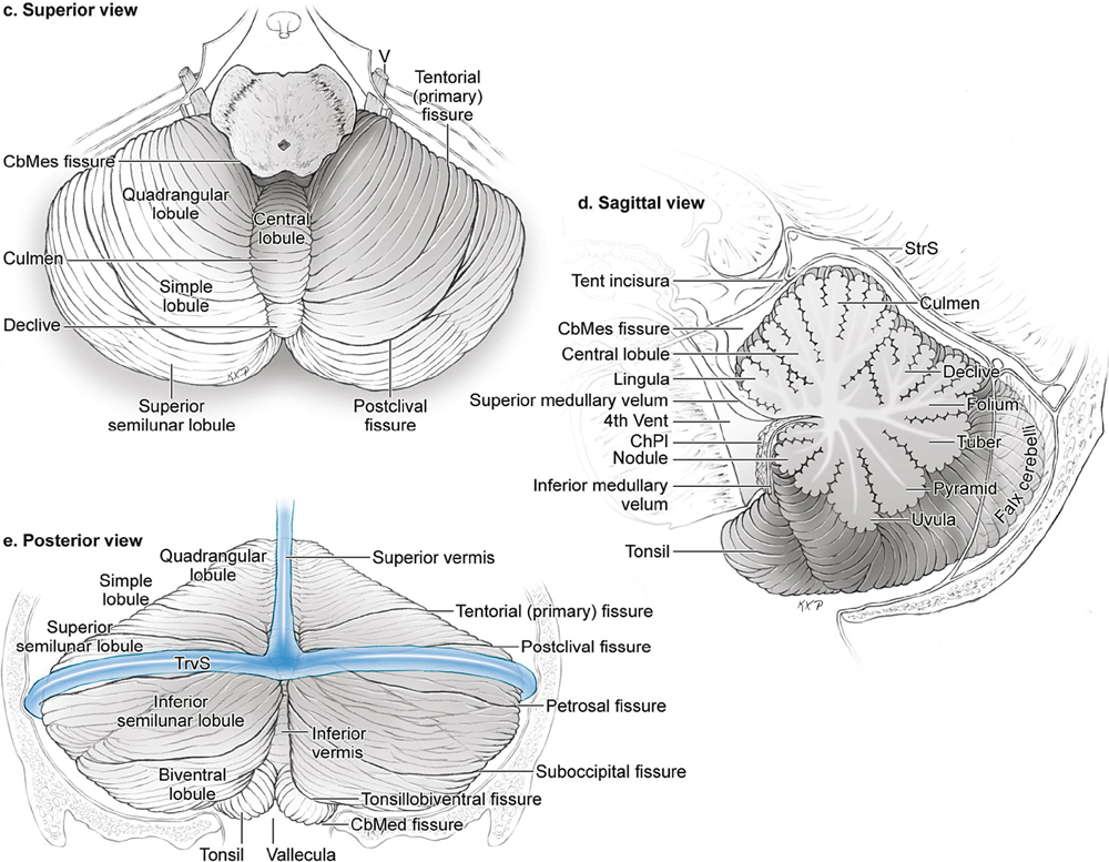
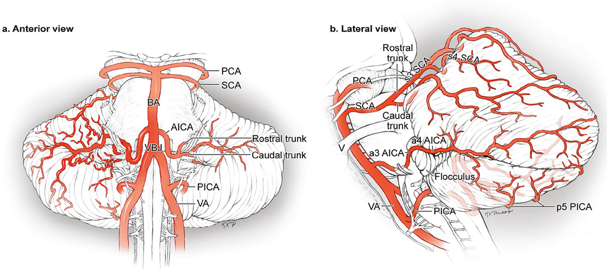
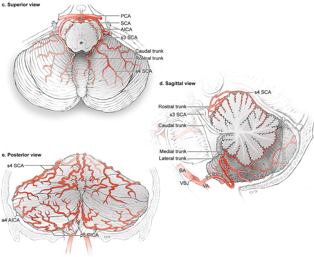
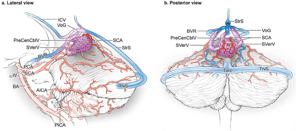
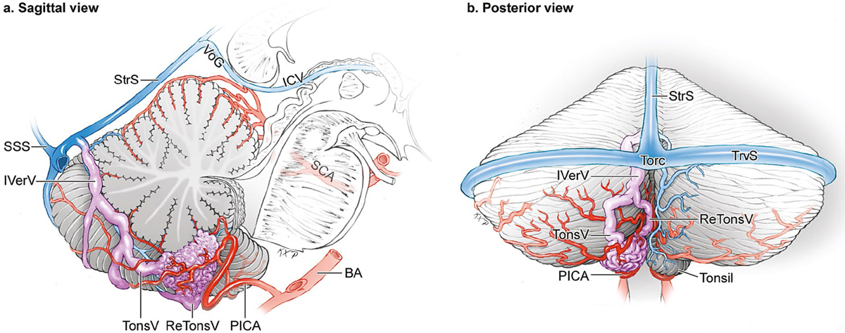
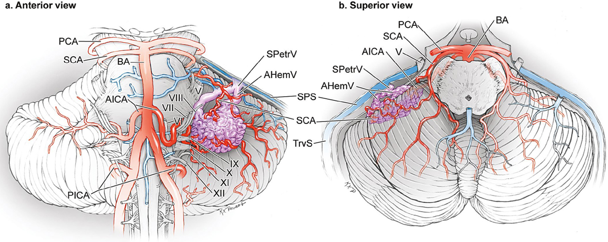
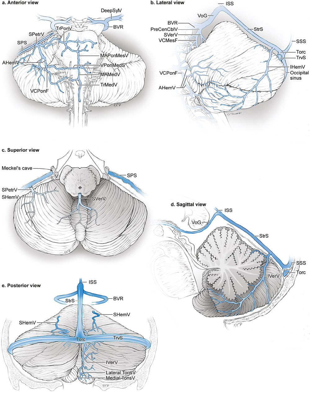
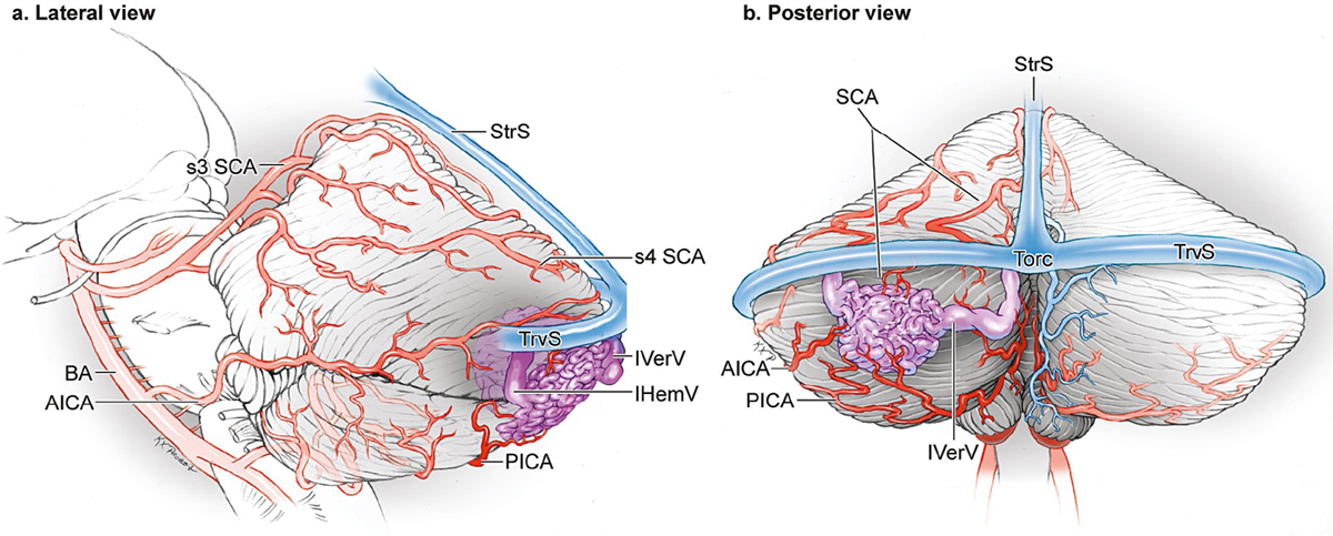
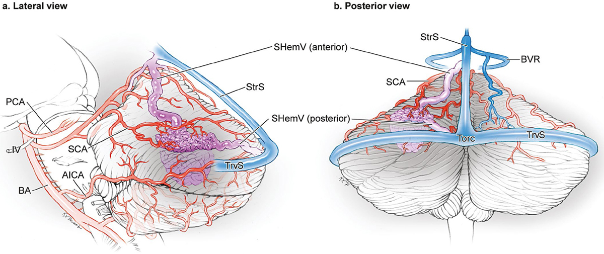
17 Cerebellar Arteriovenous Malformations The cerebellum is composed of two hemispheres joined in the midline by the vermis (Fig. 17.1). The cerebellum has three surfaces: suboccipital, tentorial, and petrosal. Each of the surfaces is divided by a major fissure named according to its surface: suboccipital, tentorial, and petrosal fissures. The hemispheric part of the tentorial surface in composed of the quadrangular, simple, and superior semilunar lobules. The tentorial surface is divided into anterior and posterior parts by its major fissure, the tentorial (primary) fissure. This fissure separates the quadrangular and simple lobules on the hemisphere, and the culmen and declive on the vermis. The postclival fissure separates the simple and superior semilunar lobules. The hemispheric portion of the suboccipital surface is composed of the superior semilunar lobule, inferior semilunar lobule, biventral lobule, and tonsil. The suboccipital surface is divided into superior and inferior parts by its major fissure, the suboccipital fissure. The vermian part of this fissure is called the prepyramidal fissure, and the hemispheric part of this fissure is called the prebiventral fissure. Minor fissures on the suboccipital surface include the petrosal (horizontal) fissure between the superior and inferior semilunar lobules, which is an extension of the major fissure on the petrosal surface, and the tonsillobiventral fissure. The inferior vermis lies in a deep vertical depression in the suboccipital surface called the posterior cerebellar incisura, which also contains the falx cerebelli. The inferior vermis forms the posterior cortical surface in the midline within this incisura. In contrast, the superior vermis is the highest point on the cerebellum, occupying the space under the straight sinus where the tentorial leaflets intersect with the falx cerebri. The superior vermis slopes downward from its apex anteriorly to the posterior cerebellar incisura. The tentorial part of the vermian surface includes (from anterior to posterior) the culmen, declive, and folium. The suboccipital part of the vermian surface includes (from superior to inferior) the tuber, pyramid, uvula, and nodule. The nodule is hidden deep to the uvula. Fig. 17.1 Microsurgical anatomy of the cerebellum. The cerebellum is composed of two hemispheres joined in the midline by the vermis and has three surfaces (suboccipital, tentorial, and petrosal), each divided by a major fissure named according to its surface (suboccipital, tentorial, and petrosal fissures). (a) Anterior view (petrosal surface). (b) Lateral view (tentorial, suboccipital, and petrosal surfaces). (c) Superior view (tentorial surface). (d) Sagittal cross-sectional view (tentorial and suboccipital surfaces of vermis). (e) Posterior view (tentorial and suboccipital surfaces). The petrosal surface of the cerebellum faces the posterior petrous bone and is divided into superior and inferior parts by the petrosal (horizontal) fissure, which extends onto the suboccipital surface between the superior and inferior semilunar lobules. The cerebellopontine angle is the V-shaped intersection of the petrosal and cerebellopontine fissures, the latter one formed where the hemispheric lobules wrap around the pons and middle cerebellar peduncle. The superior and inferior limbs of the cerebellopontine fissure meet laterally at the apex of the cerebellopontine angle, at the anterior end of the petrosal fissure. The petrosal surface is formed by the anterior surfaces of the quadrangular, simple, semilunar, and biventral lobules, and the flocculi. The two halves of the petrosal surface are not connected by a continuous strip of vermis like the other two surfaces, because of the interposition of the fourth ventricle. Anterior vermis above the fourth ventricle are the lingula, central lobule, and culmen, and below are the nodule and uvula. The arteries that supply cerebellar arteriovenous malformations (AVMs) are the cortical segments of the cerebellar arteries discussed with brainstem AVMs: the s4 superior cerebellar artery (SCA), a4 anterior inferior cerebellar artery (AICA), and the p4 and p5 posterior inferior cerebellar artery (PICA) (Fig. 17.2). The s3 segment of the SCA climbs up from the cerebellomesencephalic fissure to reach the anterior edge of the tentorial cerebellar surface, by which time it bifurcates into rostral and caudal trunks. The rostral trunk supplies the superior vermis and the caudal trunk supplies the hemispheric surface. Before exiting the cerebellomesencephalic fissure and becoming cortical segments, these trunks (particularly the rostral trunk) give rise to as many as eight precerebellar arteries that supply the dentate and deep cerebellar nuclei, and become the deep perforating artery supply to large cerebellar AVMs, or the equivalent of the “red devils” in the cerebellum that can be difficult to coagulate along the deep plane. The cortical branches divide into the hemispheric (medial, intermediate, and lateral) and vermian (medial and paramedian) segments. The AICA courses through the cerebellopontine angle near cranial nerves (CN) 7/8 and, after sending branches to the nerves entering the acoustic meatus and to the choroid plexus protruding from the foramen of Luschka, it supplies the petrosal surface of the cerebellum. The AICA typically bifurcates near the nerve complex into rostral and caudal trunks, the former supplying the middle cerebellar peduncle and superior lip of the cerebellopontine fissure, and the latter supplying the inferior lip of the cerebellopontine fissure, flocculus, and choroid plexus. Important AICA branches include brainstem perforators, labyrinthine, recurrent perforating, and subarcuate arteries. The PICA is intimate with the cerebellomedullary fissure, lower half of the ventricular roof, inferior cerebellar peduncle, and suboccipital surface. After exiting the cerebellomedullary fissure, the PICA typically bifurcates along its p4 telovelotonsillar segment into a smaller medial trunk that sends branches to the inferior vermis and adjacent hemisphere (median and paramedian vermian arteries), and a larger lateral trunk that sends branches to the suboccipital surface and tonsils (medial, intermediate, and lateral hemispheric arteries; tonsillar arteries). Cortical branches emerge from the telovelotonsillar or tonsillobiventral fissure and radiate outward from the superior and lateral borders of the tonsil. Cortical branches arising near the superior tonsil send branches to the dentate nucleus and can also be a source of deep perforating artery supply to large AVMs. Fig. 17.2 Microsurgical anatomy of the cerebellar arteries. (a) The petrosal surface of cerebellum is supplied by the cortical branches of the AICA (anterior view). (b) The lateral hemisphere is supplied by all three cerebellar arteries (lateral view). (c) The tentorial surface is supplied by cortical branches of the SCA (superior view). (d) Vermis is supplied by the SCA superiorly and the PICA inferiorly (sagittal cross-sectional view). (e) The posterior cerebellar hemispheres are supplied by the SCA superiorly, AICA laterally, and PICA inferiorly (posterior view). Superficial veins draining the tentorial surface include the superior hemispheric vein (SHemV) and superior vermian vein (SVerV) (Fig. 17.3). The suboccipital surface is drained by inferior hemispheric veins (IHemV) and inferior vermian veins (IVerV). The tonsils are drained by medial and lateral tonsillar veins (TonsV) and retrotonsillar veins (ReTonsV). The petrosal surface is drained by anterior hemispheric veins (AHemV). Deep veins course through the main fissures of the cerebellum and brainstem, including the vein of the cerebellomesencephalic fissure (VCMesF) related to the superior half of the fourth ventricular roof, the vein of the cerebellomedullary fissure (VCMedF) related to the inferior half of the fourth ventricular roof, and the vein of the cerebellopontine fissure (VCPonF) related to the lateral recess. Bridging veins from the three cerebellar surfaces include the Galenic group from superior surfaces to the vein of Galen (VoG); the tentorial group from the tentorial surface to the torcula (Torc) or tentorial sinus (TentS); and the petrosal group from the petrosal surface to the superior petrosal sinus (SPS) and inferior petrosal sinus (IPS). Fig. 17.3 Microsurgical anatomy of the cerebellar veins. (a) Anterior view (petrosal surface). (b) Lateral view (tentorial, suboccipital, and petrosal surfaces). (c) Superior view (tentorial surface). (d) Sagittal cross-sectional view (tentorial and suboccipital surfaces of vermis). (e) Posterior view (tentorial and suboccipital surfaces). Fig. 17.4 The suboccipital cerebellar AVM: (a) lateral and (b) posterior views. This AVM is based unilaterally on the suboccipital surface, can be supplied by all three cerebellar arteries, and has superficial, ascending venous drainage to the torcula and transverse sinuses. Suboccipital AVMs are based on the suboccipital cerebellar surface facing the occipital bone in the superior semilunar, inferior semilunar, and biventral lobules, below and between the transverse (TrvS) and the sigmoid sinuses (SigmS) (Fig. 17.4). They are the second most common cerebellar AVMs, accounting for one fifth of all cerebellar AVMs. They are unilateral and their location varies in the mediallateral and superior-inferior directions. Suboccipital cerebellar AVMs receive supply from the cortical branches of all three cerebellar arteries because of collateral connections on this surface, with increased participation from the SCA, AICA, and PICA with AVMs located more superiorly, laterally, and inferiorly, respectively. Venous drainage is superficial through IHemV and/or IVerV, which a bridge to Torc and TrvS. Sub-occipital AVMs are eloquent only when they extend to deep cerebellar nuclei. Midline AVMs based in the inferior vermis differ from lateral suboccipital AVMs and are categorized instead as vermian cerebellar AVMs. Tentorial AVMs are based on the tentorial surface in the quadrangular, simple, and superior semilunar lobules (Fig. 17.5). These AVMs also accounted for one fifth of all cerebellar AVMs and are unilateral. Their arterial supply is from hemispheric branches of the SCA that intersect the anterior and medial margins of the nidus. Venous drainage is through SHemV that can bridge anteriorly to the VoG, posteriorly to Torc, or superiorly to TentS or StrS. Tentorial AVMs typically do not extend to the deep cerebellar nuclei and are non-eloquent. Midline AVMs based in the superior vermis differ from tentorial AVMs and are categorized instead as vermian AVMs. Vermian AVMs are the most common cerebellar AVM (30%) and sit in the midline in the superior vermis (tentorial surface, Fig. 17.6) or the inferior vermis (suboccipital surface), with the superior vermian being more common (90% vs. 10%, respectively). Superior vermian AVMs are located in the lingula, central lobule, culmen, and/or declive, whereas inferior vermian AVMs are located in the folium, tuber, pyramid, uvula, and/or nodule. Arterial supply is bilateral and depends on location, with superior vermian AVMs fed by the SCAs and inferior vermian AVMs fed by PICAs. Superior vermian AVMs drain deep to SVerV and VoG, whereas inferior vermian AVMs drain superficially to IVerV and Torc. Most vermian AVMs are non-eloquent. Fig. 17.5 The tentorial cerebellar AVM: (a) lateral and (b) posterior views. This AVM is based unilaterally on the tentorial surface, is supplied by hemispheric branches of the SCA, and drains to the superior hemispheric veins that may course deeply to the VoG (anterior SHemV) or superficially to the torcula or transverse sinus (posterior SHemV). Fig. 17.6 The vermian cerebellar AVM: (a) lateral and (b) posterior views. This AVM is based in the midline either on the superior vermis (tentorial surface, as shown) or on the inferior vermis (suboccipital surface, not shown). This AVM is supplied bilaterally by the SCA in the superior vermis and by the PICA in the inferior vermis. Superior vermian AVMs drain deeply to the VoG via SVerV and inferior vermian AVMs drain superficially to the torcula via IVerV. Tonsillar AVMs lie in the cerebellar tonsils at the lower edge of the suboccipital surface (Fig. 17.7) and were the least common cerebellar AVM (8%). These are unilateral AVMs fed by the ipsilateral PICA, and have superficial drainage to medial and lateral TonsV as well as ReTonsV. Tonsillar AVMs are small in size and non-eloquent. Fig. 17.7 The tonsillar cerebellar AVM: (a) sagittal cross-sectional and (b) posterior views. This AVM is based in the cerebellar tonsil unilaterally, supplied by the ipsilateral PICA, and drained by the tonsillar veins. Petrosal AVMs are based on the petrosal surface, which is the anterior cerebellum that faces the posterior petrous bone (Fig. 17.8). These AVMs are located in the parts of the cerebellar lobules that wrap around to the anterior surface: the quadrangular, simple, superior semilunar, inferior semilunar, and biventral lobules. They are distinct from lateral pontine AVMs that are based in the lateral pons and middle cerebellar peduncle. These AVMs are unilateral and supplied by cortical branches of the AICA. They drain to AHemV and VCPonF, which then course to SPetrV and SPS. These AVMs are non-eloquent, but not far from CN5, CN7/8, and pons. Fig. 17.8 The petrosal cerebellar AVM: (a) superior and (b) anterior views. This AVM is based on the petrosal surface lateral to the brachium pontis. It is supplied by cortical branches of the AICA and is drained by the anterior hemispheric veins and petrosal vein.
 Microsurgical Anatomy
Brain
Arteries
Veins
 Five Cerebellar AVM Subtypes
The Suboccipital Cerebellar AVM
The Tentorial Cerebellar AVM
The Vermian Cerebellar AVM
The Tonsillar Cerebellar AVM
The Petrosal Cerebellar AVM
Related posts:


Stay updated, free articles. Join our Telegram channel

Full access? Get Clinical Tree


